Copyright
©The Author(s) 2021.
World J Clin Cases. Sep 6, 2021; 9(25): 7579-7587
Published online Sep 6, 2021. doi: 10.12998/wjcc.v9.i25.7579
Published online Sep 6, 2021. doi: 10.12998/wjcc.v9.i25.7579
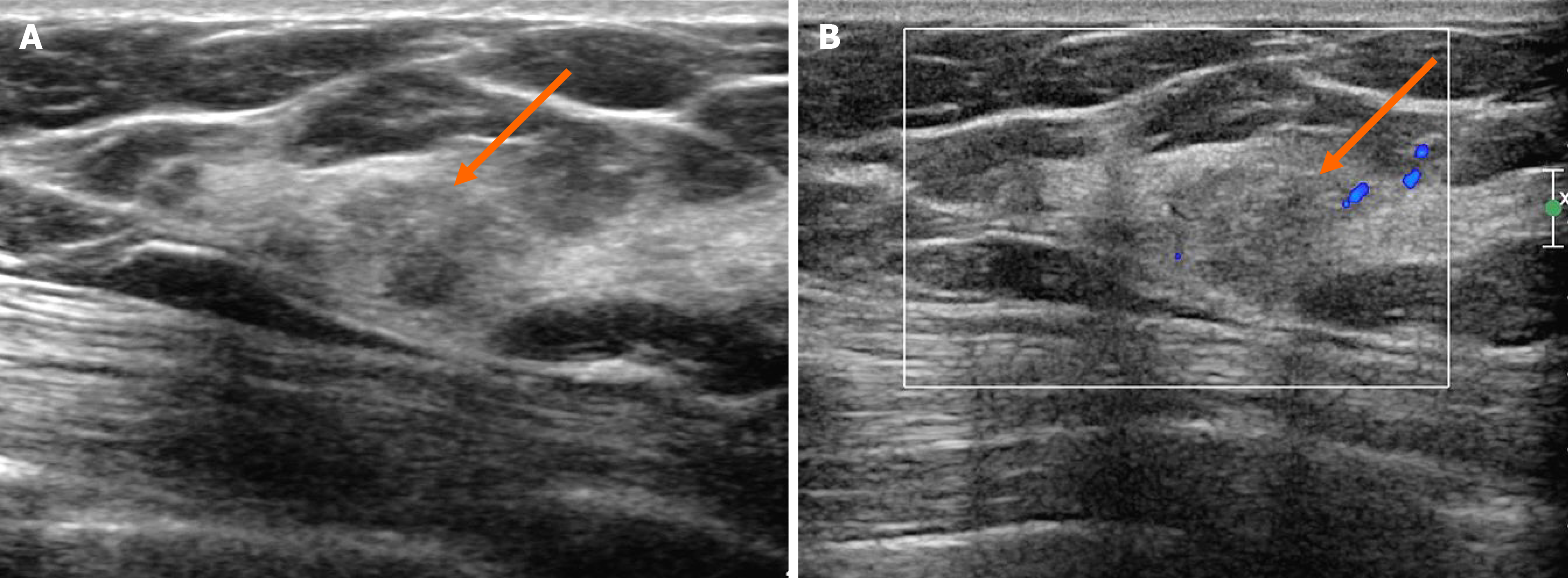
Figure 2 Ultrasonography showing an irregular mass.
A: On B-mode ultrasonography, the lesion showed an irregular shape and angular margin (arrow). The echogenicity was slightly higher than that of fat; B: In the color Doppler study, there was no internal or rim vascularity (arrow).
- Citation: An JK, Woo JJ, Kim EK, Kwak HY. Breast adenoid cystic carcinoma arising in microglandular adenosis: A case report and review of literature. World J Clin Cases 2021; 9(25): 7579-7587
- URL: https://www.wjgnet.com/2307-8960/full/v9/i25/7579.htm
- DOI: https://dx.doi.org/10.12998/wjcc.v9.i25.7579


