Copyright
©The Author(s) 2021.
World J Clin Cases. Sep 6, 2021; 9(25): 7372-7380
Published online Sep 6, 2021. doi: 10.12998/wjcc.v9.i25.7372
Published online Sep 6, 2021. doi: 10.12998/wjcc.v9.i25.7372
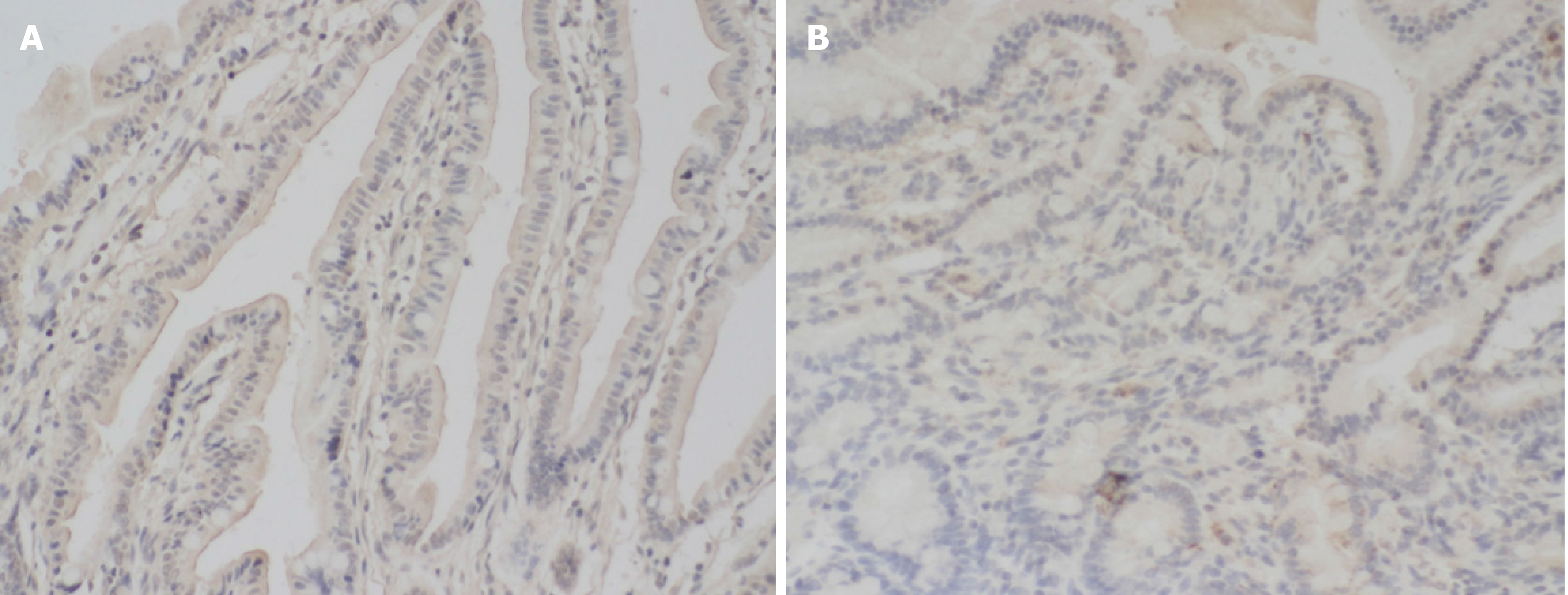
Figure 2 Mucin 1 protein immunohistochemical staining.
A: The intestinal mucosal tissue of the control group; B: The necrotizing enterocolitis intestinal mucosal tissue. It can be seen that the coloring is deeper in the control group, and the mucin 1 protein expression intensity is stronger, × 200.
- Citation: Pan HX, Zhang CS, Lin CH, Chen MM, Zhang XZ, Yu N. Mucin 1 and interleukin-11 protein expression and inflammatory reactions in the intestinal mucosa of necrotizing enterocolitis children after surgery. World J Clin Cases 2021; 9(25): 7372-7380
- URL: https://www.wjgnet.com/2307-8960/full/v9/i25/7372.htm
- DOI: https://dx.doi.org/10.12998/wjcc.v9.i25.7372


