Copyright
©The Author(s) 2021.
World J Clin Cases. Aug 16, 2021; 9(23): 6935-6942
Published online Aug 16, 2021. doi: 10.12998/wjcc.v9.i23.6935
Published online Aug 16, 2021. doi: 10.12998/wjcc.v9.i23.6935
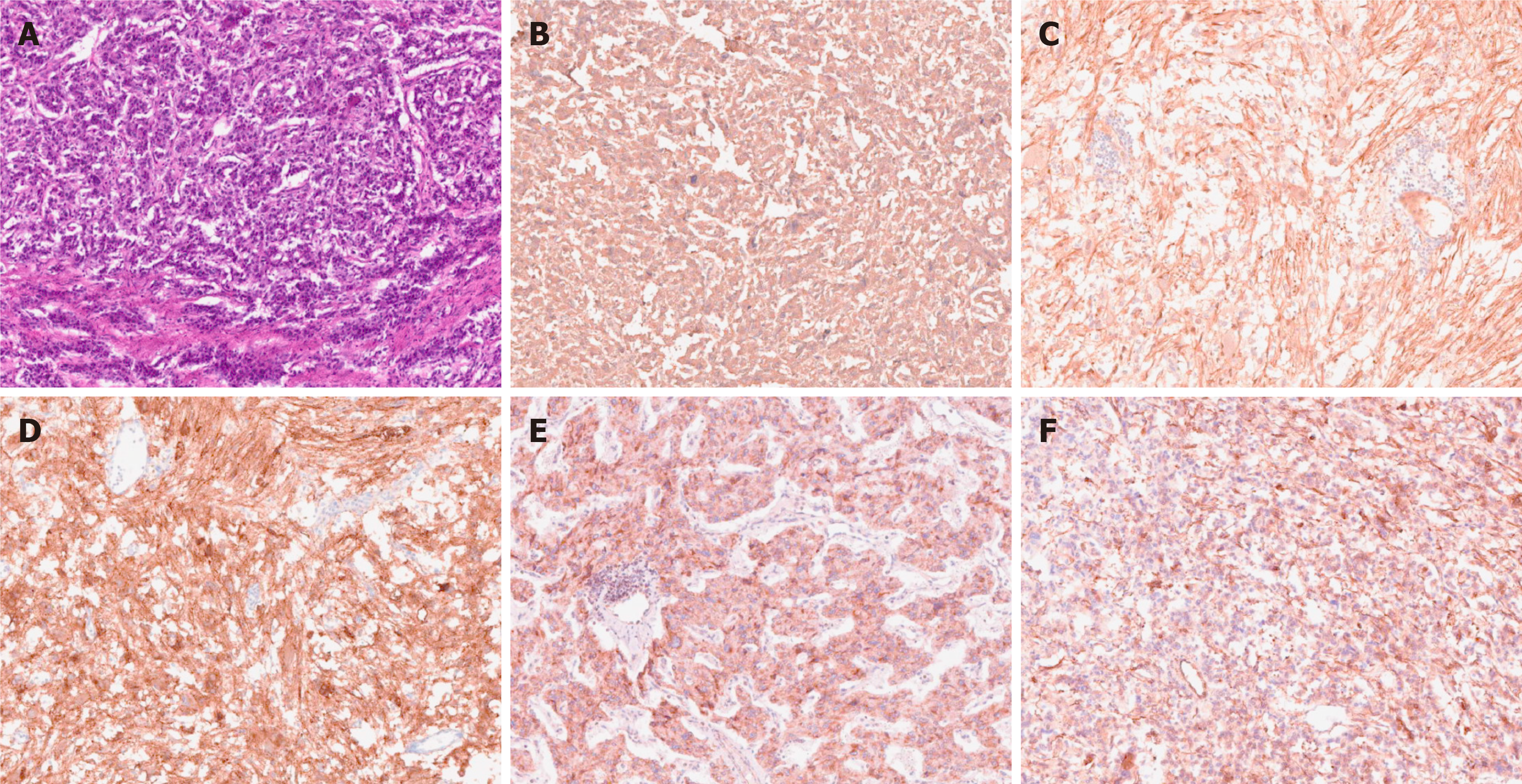
Figure 6 Pathological photos.
Histological examination: A: HE staining; Immunohistochemistry: B: Chromogranin A (+); C: Soluble protein-100 (+); D: Synaptophysin (+); E: Succinate dehydrogenase B (+); F: Neuron specific enolase (+).
- Citation: Liu C, Wen J, Li HZ, Ji ZG. Combined thoracoscopic and laparoscopic approach to remove a large retroperitoneal compound paraganglioma: A case report. World J Clin Cases 2021; 9(23): 6935-6942
- URL: https://www.wjgnet.com/2307-8960/full/v9/i23/6935.htm
- DOI: https://dx.doi.org/10.12998/wjcc.v9.i23.6935


