Copyright
©The Author(s) 2021.
World J Clin Cases. Aug 16, 2021; 9(23): 6935-6942
Published online Aug 16, 2021. doi: 10.12998/wjcc.v9.i23.6935
Published online Aug 16, 2021. doi: 10.12998/wjcc.v9.i23.6935
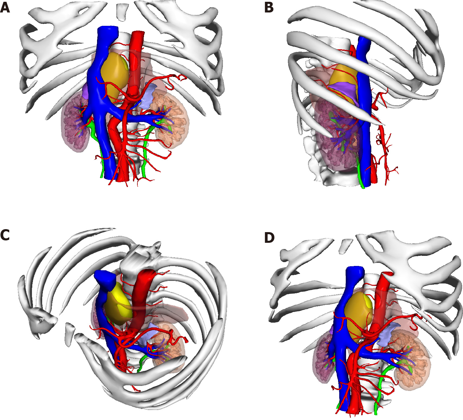
Figure 3 The position of the tumor shown in 3-dimensional printing.
A: Front view; B: Right side view; C: Diagonally above view; D: Diagonally below view. Yellow: Tumor; Orange: Muscle; Blue: Vein; Red: Artery.
- Citation: Liu C, Wen J, Li HZ, Ji ZG. Combined thoracoscopic and laparoscopic approach to remove a large retroperitoneal compound paraganglioma: A case report. World J Clin Cases 2021; 9(23): 6935-6942
- URL: https://www.wjgnet.com/2307-8960/full/v9/i23/6935.htm
- DOI: https://dx.doi.org/10.12998/wjcc.v9.i23.6935


