Copyright
©The Author(s) 2021.
World J Clin Cases. Aug 16, 2021; 9(23): 6935-6942
Published online Aug 16, 2021. doi: 10.12998/wjcc.v9.i23.6935
Published online Aug 16, 2021. doi: 10.12998/wjcc.v9.i23.6935
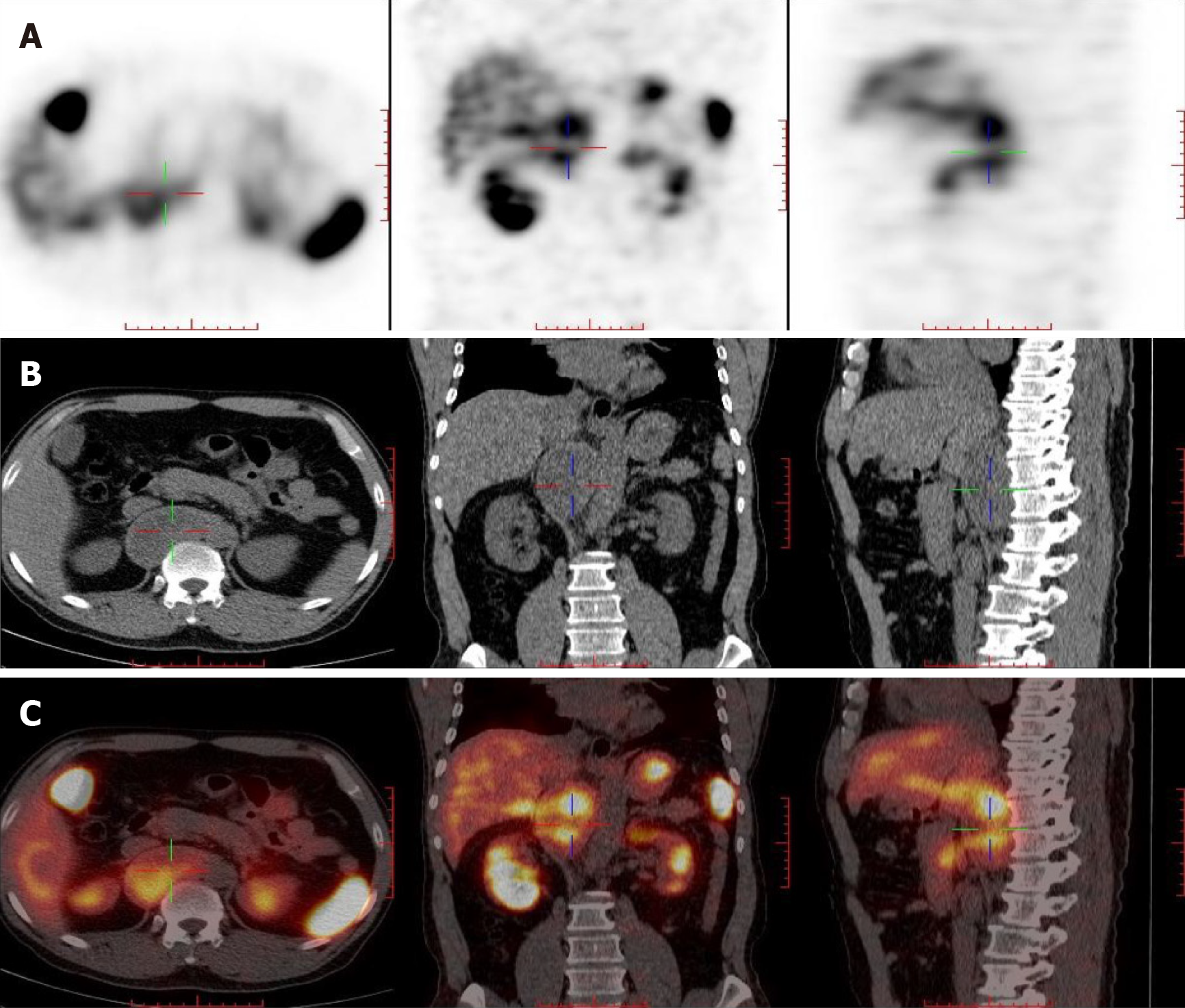
Figure 2 Somatostatin receptor imaging.
A: Contrast agent imaging; B: Computed tomography; C: Fusion imaging of A and B. At the T10-L1 vertebral level on the right side of the abdominal aorta and behind the inferior vena cava, there is a cystic solid space, measuring 7.3 cm × 3.6 cm × 9.4 cm in size, with increased radioactive uptake.
- Citation: Liu C, Wen J, Li HZ, Ji ZG. Combined thoracoscopic and laparoscopic approach to remove a large retroperitoneal compound paraganglioma: A case report. World J Clin Cases 2021; 9(23): 6935-6942
- URL: https://www.wjgnet.com/2307-8960/full/v9/i23/6935.htm
- DOI: https://dx.doi.org/10.12998/wjcc.v9.i23.6935


