Copyright
©The Author(s) 2021.
World J Clin Cases. Aug 16, 2021; 9(23): 6935-6942
Published online Aug 16, 2021. doi: 10.12998/wjcc.v9.i23.6935
Published online Aug 16, 2021. doi: 10.12998/wjcc.v9.i23.6935
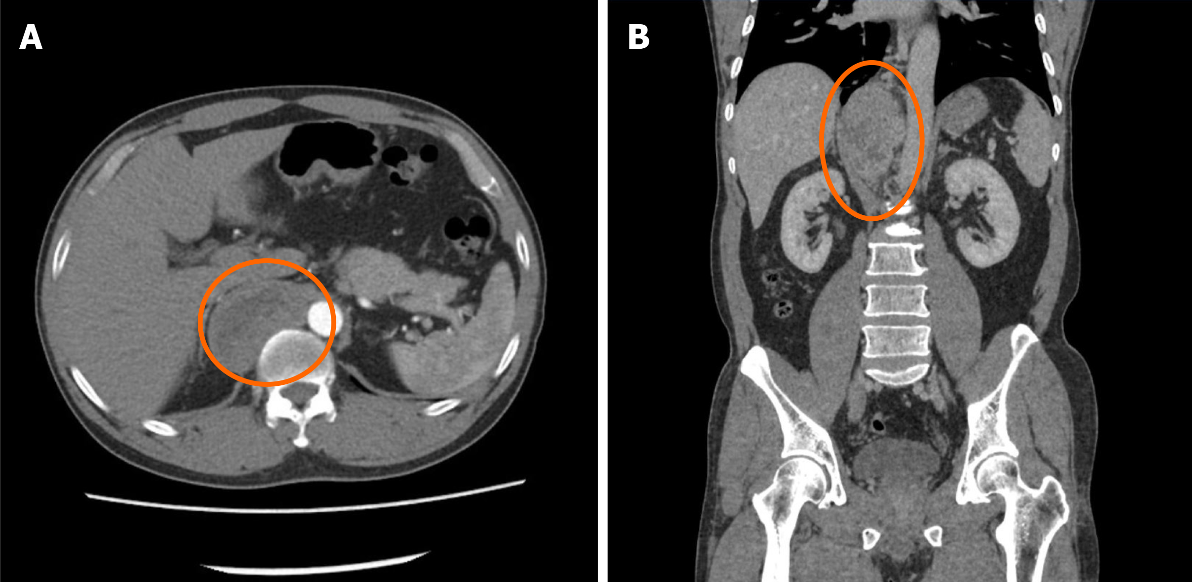
Figure 1 Contrast-enhanced computed tomography + three-dimensional reconstruction of the abdomen and pelvis.
A: Contrast-enhanced computed tomography; B: Three-dimensional reconstruction. At the T10-L1 level of the posterior mediastinal spine on the right anterior side (posterior space of the right phrenic foot), there is an elliptical mixed density shadow, with smooth borders, and the size is about 3.3 cm × 6.9 cm × 8.4 cm.
- Citation: Liu C, Wen J, Li HZ, Ji ZG. Combined thoracoscopic and laparoscopic approach to remove a large retroperitoneal compound paraganglioma: A case report. World J Clin Cases 2021; 9(23): 6935-6942
- URL: https://www.wjgnet.com/2307-8960/full/v9/i23/6935.htm
- DOI: https://dx.doi.org/10.12998/wjcc.v9.i23.6935


