
















©The Author(s) 2021.
World J Clin Cases. Aug 16, 2021; 9(23): 6872-6878
Published online Aug 16, 2021. doi: 10.12998/wjcc.v9.i23.6872
Published online Aug 16, 2021. doi: 10.12998/wjcc.v9.i23.6872
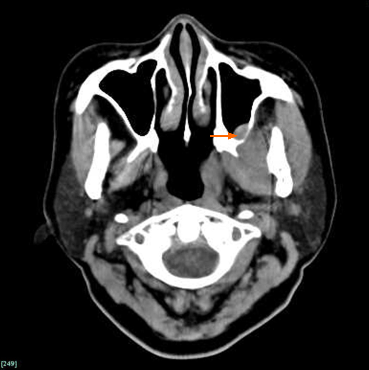
Figure 1 Obvious enlargement of the left side pterygoid muscles appear on computed tomography scanning.
The boundary of the lateral and medial pterygoid muscles is obscure. Bone destruction and thickened mucous membrane on the maxilla sinus back wall appear as well (arrow).
- Citation: Bi L, Wei D, Wang B, He JF, Zhu HY, Wang HM. Trismus originating from rare fungal myositis in pterygoid muscles: A case report. World J Clin Cases 2021; 9(23): 6872-6878
- URL: https://www.wjgnet.com/2307-8960/full/v9/i23/6872.htm
- DOI: https://dx.doi.org/10.12998/wjcc.v9.i23.6872