Copyright
©The Author(s) 2021.
World J Clin Cases. Aug 16, 2021; 9(23): 6846-6857
Published online Aug 16, 2021. doi: 10.12998/wjcc.v9.i23.6846
Published online Aug 16, 2021. doi: 10.12998/wjcc.v9.i23.6846
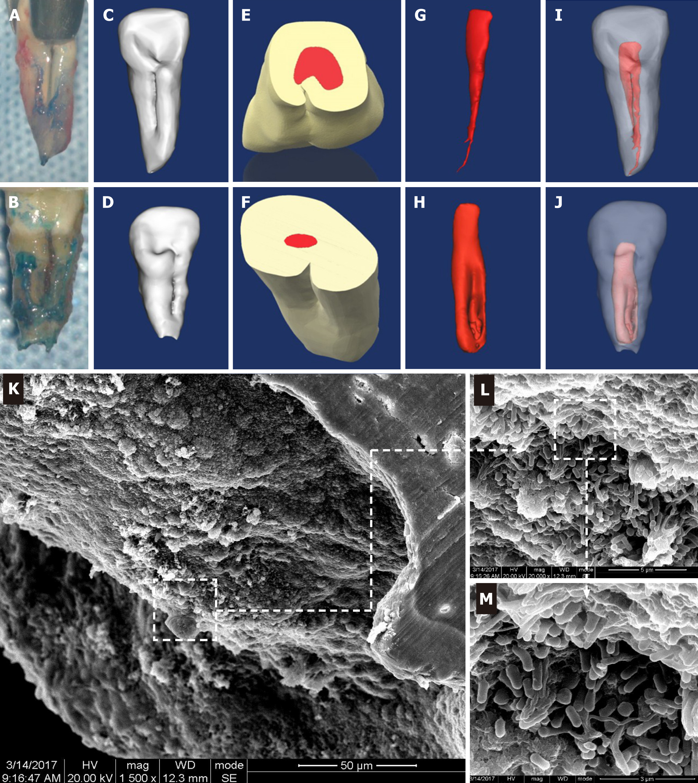
Figure 1 Three-dimensional reconstruction of the teeth with palatal radicular groove and representative scanning electron microscope images for resected root apex.
A and B: Intraoperative awareness of palatal groove during intentional replantation; C and D: Lingual view of the three-dimensional reconstruction models of type I and type II palatal grooves; E-J: Shapes of the pulp cavity associated with type I and II palatal grooves and their spatial and configurational relationship with the root; K-M: Gradually magnified scanning electron microscope photograph series of the biofilm covering the root apex of tooth with palatal radicular groove associated infection (K: × 1500; L: × 20000; M: × 40000).
- Citation: Tan XL, Chen X, Fu YJ, Ye L, Zhang L, Huang DM. Diverse microbiota in palatal radicular groove analyzed by Illumina sequencing: Four case reports. World J Clin Cases 2021; 9(23): 6846-6857
- URL: https://www.wjgnet.com/2307-8960/full/v9/i23/6846.htm
- DOI: https://dx.doi.org/10.12998/wjcc.v9.i23.6846


