
















©The Author(s) 2021.
World J Clin Cases. Aug 16, 2021; 9(23): 6824-6831
Published online Aug 16, 2021. doi: 10.12998/wjcc.v9.i23.6824
Published online Aug 16, 2021. doi: 10.12998/wjcc.v9.i23.6824
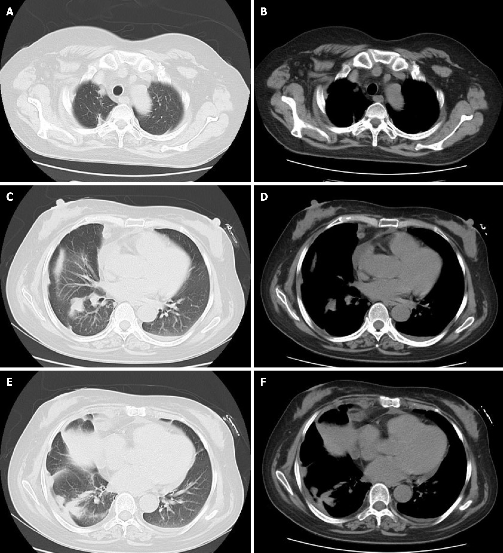
Figure 5 Axial computed tomography showed that the lesions were obviously improved in terms of absorption after 1 mo of treatment.
A and B: At the upper level of the lung, the lesions located in the right pleural and mediastinum areas were significantly reduced; C and D: At the middle level of the lung, the lesions located in the right pleura, mediastinum, and interlobar fissure areas were significantly reduced; E and F: At the lower level of the lung, the lesions located in the right pleura and mediastinum areas were significantly reduced and atelectasis of adjacent lung was markedly improved.
- Citation: Wang P, Yi ML, Zhang CZ. Nocardiosis with diffuse involvement of the pleura: A case report. World J Clin Cases 2021; 9(23): 6824-6831
- URL: https://www.wjgnet.com/2307-8960/full/v9/i23/6824.htm
- DOI: https://dx.doi.org/10.12998/wjcc.v9.i23.6824