
















©The Author(s) 2021.
World J Clin Cases. Aug 16, 2021; 9(23): 6781-6788
Published online Aug 16, 2021. doi: 10.12998/wjcc.v9.i23.6781
Published online Aug 16, 2021. doi: 10.12998/wjcc.v9.i23.6781
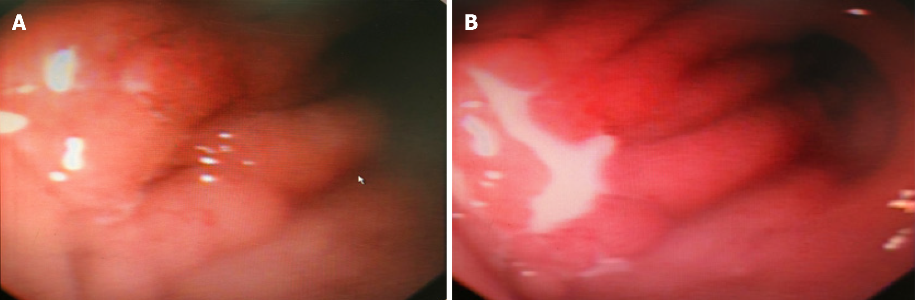
Figure 4 Gastroscopic view.
A: The mucosa of the anterior wall of the duodenal bulb is shown to be rough and uneven, with small bubbles throughout; B: Milky white pus is seen exudating from the anterior wall of the duodenal bulb, and the foreign body was considered to have punctured the anterior wall of the duodenal bulb.
- Citation: Pan W, Lin LJ, Meng ZW, Cai XR, Chen YL. Hepatic abscess caused by esophageal foreign body misdiagnosed as cystadenocarcinoma by magnetic resonance imaging: A case report. World J Clin Cases 2021; 9(23): 6781-6788
- URL: https://www.wjgnet.com/2307-8960/full/v9/i23/6781.htm
- DOI: https://dx.doi.org/10.12998/wjcc.v9.i23.6781