Copyright
©The Author(s) 2021.
World J Clin Cases. Aug 6, 2021; 9(22): 6522-6530
Published online Aug 6, 2021. doi: 10.12998/wjcc.v9.i22.6522
Published online Aug 6, 2021. doi: 10.12998/wjcc.v9.i22.6522
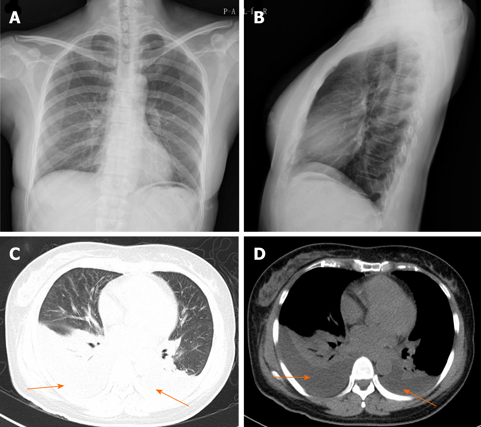
Figure 1 Preoperative chest X-ray films and chest computed tomography of postoperative day 2.
A and B: Preoperative frontal (A) and lateral (B) chest films show a normal chest; C and D: Postoperative high-resolution computed tomography shows bilateral small to moderate effusion (orange arrow).
- Citation: Huang XM, Luo M, Ran LY, You XH, Wu DW, Huang SS, Gong Q. Chylothorax following posterior low lumbar fusion surgery: A case report. World J Clin Cases 2021; 9(22): 6522-6530
- URL: https://www.wjgnet.com/2307-8960/full/v9/i22/6522.htm
- DOI: https://dx.doi.org/10.12998/wjcc.v9.i22.6522


