Copyright
©The Author(s) 2021.
World J Clin Cases. Aug 6, 2021; 9(22): 6485-6492
Published online Aug 6, 2021. doi: 10.12998/wjcc.v9.i22.6485
Published online Aug 6, 2021. doi: 10.12998/wjcc.v9.i22.6485
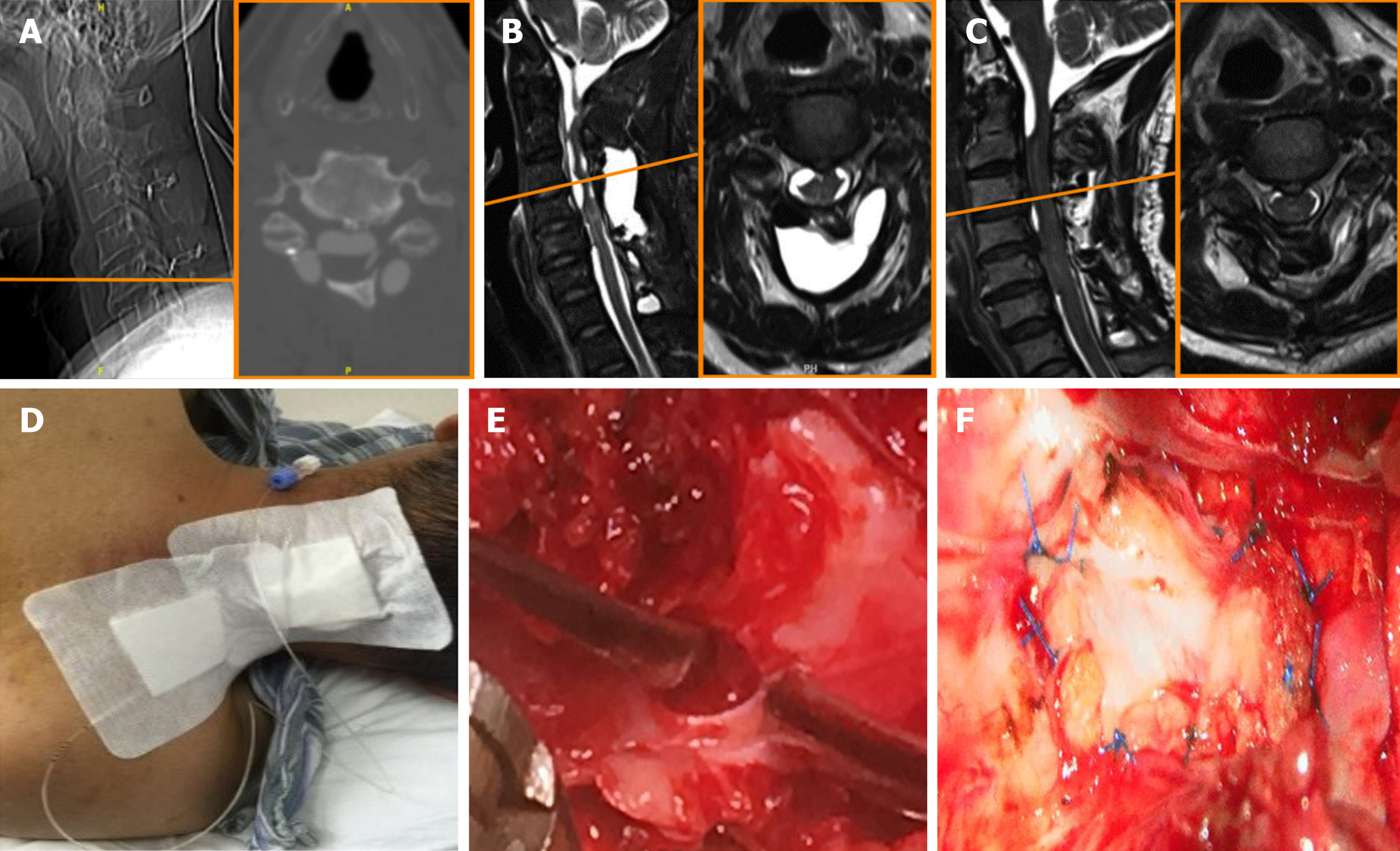
Figure 2 Imaging examination and treatment at our hospital.
A: Computed tomography myelography revealing that the defect of dural-arachnoid was located at the C5 level and close to the lower edge of the fixed plate; B: Sagittal and axial view of cervical magnetic resonance imaging before pseudomeningocele drainage; C: Sagittal and axial view after pseudomeningocele drainage revealing a significant decrease in the cystic volume; D: The patient undergoing pseudomeningocele drainage; E: Intraoperative photograph demonstrating the dural-arachnoid defect; F: Dural-arachnoid defect was repaired with autologous fascia.
- Citation: Huang HH, Cheng ZH, Ding BZ, Zhao J, Zhao CQ. Subdural fluid collection rather than meningitis contributes to hydrocephalus after cervical laminoplasty: A case report. World J Clin Cases 2021; 9(22): 6485-6492
- URL: https://www.wjgnet.com/2307-8960/full/v9/i22/6485.htm
- DOI: https://dx.doi.org/10.12998/wjcc.v9.i22.6485


