Copyright
©The Author(s) 2021.
World J Clin Cases. Aug 6, 2021; 9(22): 6469-6477
Published online Aug 6, 2021. doi: 10.12998/wjcc.v9.i22.6469
Published online Aug 6, 2021. doi: 10.12998/wjcc.v9.i22.6469
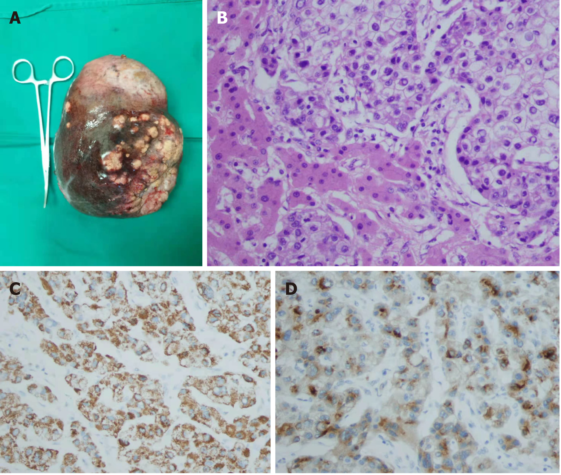
Figure 5 Resected specimen (diaphragmatic surface of liver).
A: Microscopic findings. Hematoxylin and eosin (H&E) staining showing polygonal tumor cells in a trabecular arrangement, H&E × 400; B-D: Immunohistochemistry showing a strong and diffuse cytoplasmic (B) positivity with Hep Par 1 (C) and glypican 3 (D), × 400. The tumor cells were negative for cytokeratin 7 and cytokeratin 20.
- Citation: Zhang JJ, Wang ZX, Niu JX, Zhang M, An N, Li PF, Zheng WH. Successful totally laparoscopic right trihepatectomy following conversion therapy for hepatocellular carcinoma: A case report. World J Clin Cases 2021; 9(22): 6469-6477
- URL: https://www.wjgnet.com/2307-8960/full/v9/i22/6469.htm
- DOI: https://dx.doi.org/10.12998/wjcc.v9.i22.6469


