Copyright
©The Author(s) 2021.
World J Clin Cases. Aug 6, 2021; 9(22): 6457-6463
Published online Aug 6, 2021. doi: 10.12998/wjcc.v9.i22.6457
Published online Aug 6, 2021. doi: 10.12998/wjcc.v9.i22.6457
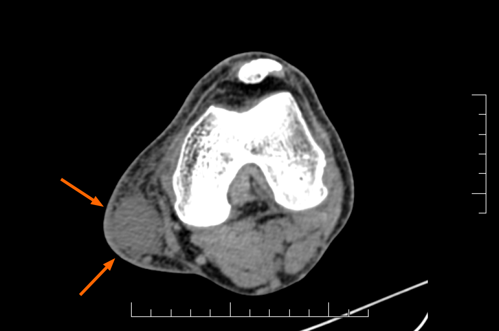
Figure 2 Axial computed tomography image.
The axial computed tomography image revealed that the oval well-defined lesion (orange arrows) on the medial side of the left knee had a lower computed tomography value (34 HU) than adjacent anatomical structures (62 HU), which were compressed and displaced.
- Citation: Yang CM, Li JM, Wang R, Lu LG. Malignant peripheral nerve sheath tumor in an elderly patient with superficial spreading melanoma: A case report. World J Clin Cases 2021; 9(22): 6457-6463
- URL: https://www.wjgnet.com/2307-8960/full/v9/i22/6457.htm
- DOI: https://dx.doi.org/10.12998/wjcc.v9.i22.6457


