Copyright
©The Author(s) 2021.
World J Clin Cases. Jul 26, 2021; 9(21): 6145-6154
Published online Jul 26, 2021. doi: 10.12998/wjcc.v9.i21.6145
Published online Jul 26, 2021. doi: 10.12998/wjcc.v9.i21.6145
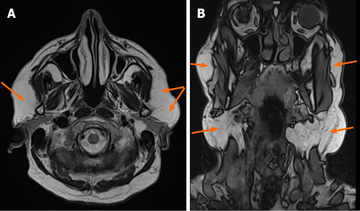
Figure 3 Head and neck magnetic resonance imaging images showing the localization of fat masses in parotid and submandibular areas.
A: Axial plane: fat deposits adjacent to parotid salivary glands (arrows); B: Coronal plane: significantly enlarged subcutaneous fat tissue, lipomatous masses below the mandible, in the upper part of the neck and in the area of parotid salivary glands (arrows).
- Citation: Seskute G, Dapkute A, Kausaite D, Strainiene S, Talijunas A, Butrimiene I. Multidisciplinary diagnostic dilemma in differentiating Madelung’s disease — the value of superb microvascular imaging technique: A case report. World J Clin Cases 2021; 9(21): 6145-6154
- URL: https://www.wjgnet.com/2307-8960/full/v9/i21/6145.htm
- DOI: https://dx.doi.org/10.12998/wjcc.v9.i21.6145


