Copyright
©The Author(s) 2021.
World J Clin Cases. Jul 26, 2021; 9(21): 6145-6154
Published online Jul 26, 2021. doi: 10.12998/wjcc.v9.i21.6145
Published online Jul 26, 2021. doi: 10.12998/wjcc.v9.i21.6145
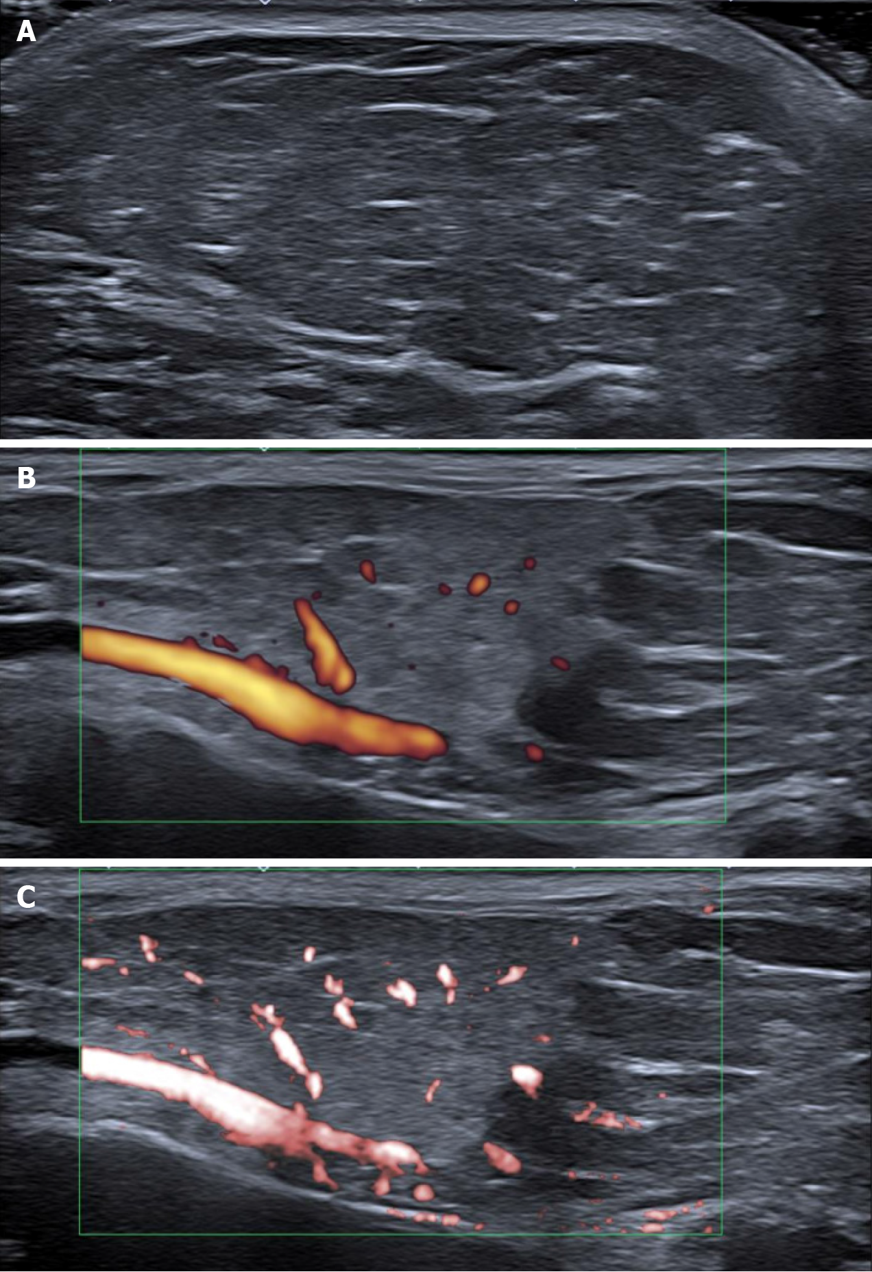
Figure 2 Ultrasound images of tumor masses in the area of the parotid gland.
A: Grayscale ultrasound showing typical superficial lipoma well-circumscribed with parallel linear and thin echogenic lines; B: Power Doppler showing several small internal dots minimal flow/vascularity; C: Superb microvascular imaging confirming low vascularity (several unrelated dots), which is a weak suspicious for liposarcoma.
- Citation: Seskute G, Dapkute A, Kausaite D, Strainiene S, Talijunas A, Butrimiene I. Multidisciplinary diagnostic dilemma in differentiating Madelung’s disease — the value of superb microvascular imaging technique: A case report. World J Clin Cases 2021; 9(21): 6145-6154
- URL: https://www.wjgnet.com/2307-8960/full/v9/i21/6145.htm
- DOI: https://dx.doi.org/10.12998/wjcc.v9.i21.6145


