Copyright
©The Author(s) 2021.
World J Clin Cases. Jul 26, 2021; 9(21): 6102-6109
Published online Jul 26, 2021. doi: 10.12998/wjcc.v9.i21.6102
Published online Jul 26, 2021. doi: 10.12998/wjcc.v9.i21.6102
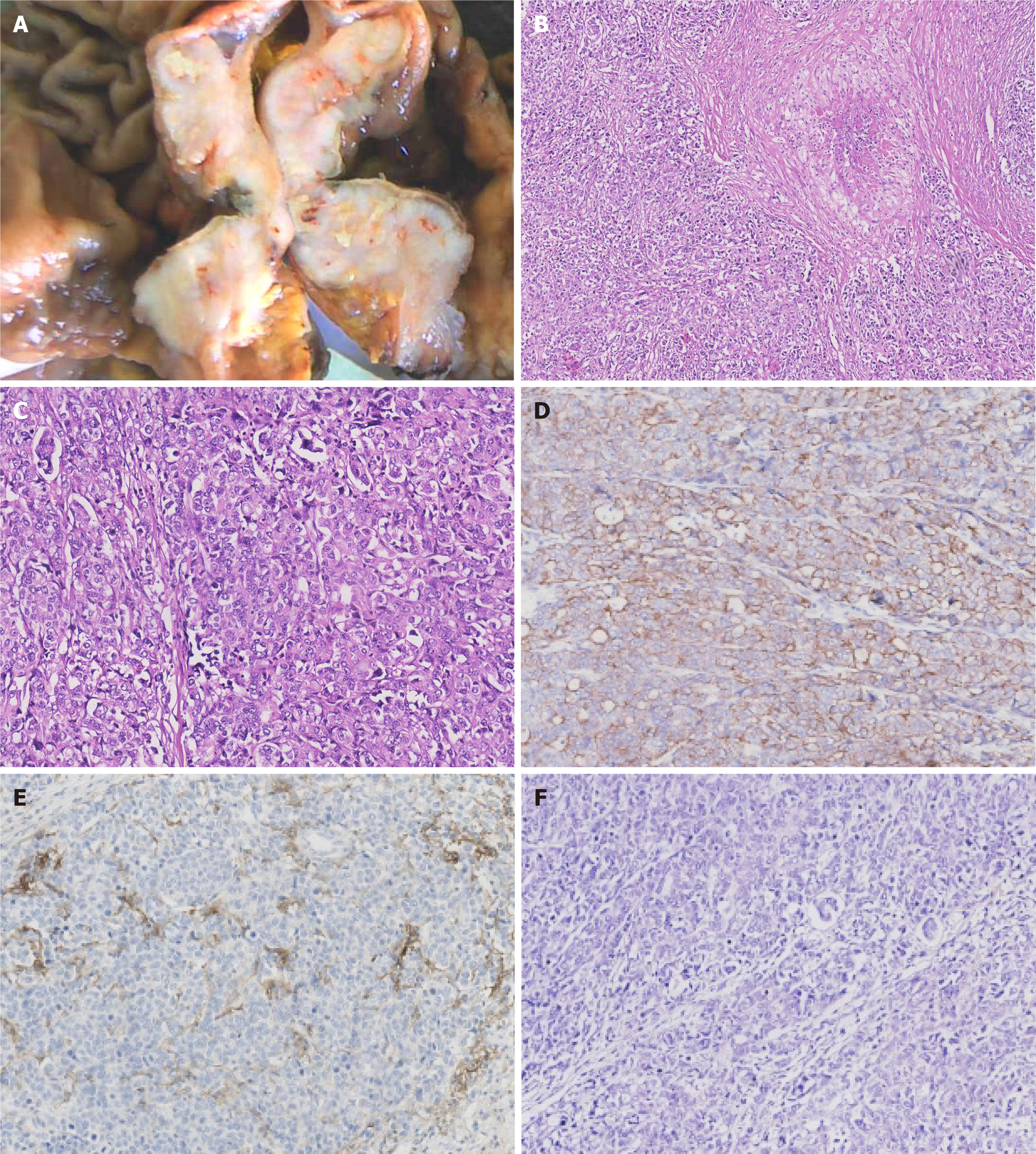
Figure 1 Pathological characteristics.
A: Gross appearance of the tumor; B: Microscopic appearance of the tumor with necrosis; C: Microscopic appearance of the tumor with solid pattern and lymphatic stroma; D: The tumor cells were positive for cytokeratin AE1/AE3 by immunohistochemical staining; E: High expression of PDL1 (22C3) CPS>1; F: Epstein–Barr virus encoded small RNA was negative (figures at 100-200 × magnification).
- Citation: Yue M, Liu JY, Liu YP. Unusual immunohistochemical “null” pattern of four mismatch repair proteins in gastric cancer: A case report. World J Clin Cases 2021; 9(21): 6102-6109
- URL: https://www.wjgnet.com/2307-8960/full/v9/i21/6102.htm
- DOI: https://dx.doi.org/10.12998/wjcc.v9.i21.6102


