Copyright
©The Author(s) 2021.
World J Clin Cases. Jul 26, 2021; 9(21): 6005-6008
Published online Jul 26, 2021. doi: 10.12998/wjcc.v9.i21.6005
Published online Jul 26, 2021. doi: 10.12998/wjcc.v9.i21.6005
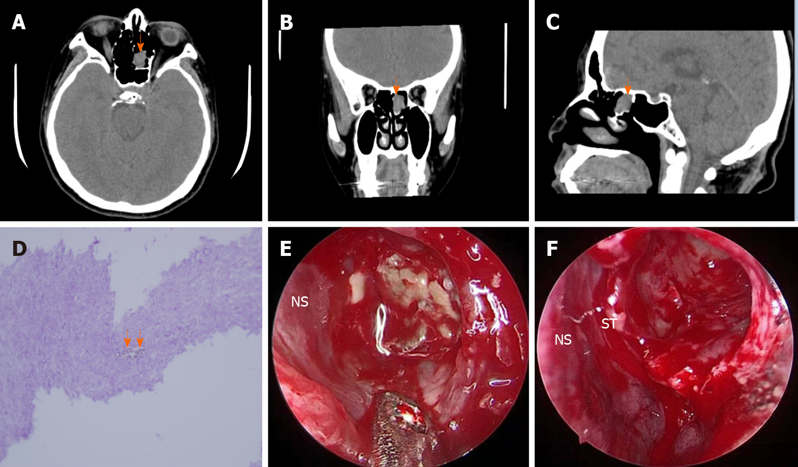
Figure 1 Computed tomography, histopathology, and nasal endoscopic images of the case.
A-C: Computed tomography (CT) scan of the nose and paranasal sinuses showed a soft-tissue density with a round surface (arrow) in the left ethmoid roof cell (axial, coronal, and sagittal CT scans); D: Histopathological image reveals fungal hyphae (arrow) (hematoxylin and eosin staining 400 ×); E: A small, dark-brownish mass wass observed in the left ethmoid sinus using a “0” nasal endoscope; F: After functional endoscopic sinus surgery, the mass was removed. NS: Nasal septum; ST: Superior turbinate.
- Citation: Zhou LQ, Li M, Li YQ, Wang YJ. Isolated fungus ball in a single cell of the left ethmoid roof: A case report. World J Clin Cases 2021; 9(21): 6005-6008
- URL: https://www.wjgnet.com/2307-8960/full/v9/i21/6005.htm
- DOI: https://dx.doi.org/10.12998/wjcc.v9.i21.6005


