
















©The Author(s) 2021.
World J Clin Cases. Jul 16, 2021; 9(20): 5631-5636
Published online Jul 16, 2021. doi: 10.12998/wjcc.v9.i20.5631
Published online Jul 16, 2021. doi: 10.12998/wjcc.v9.i20.5631
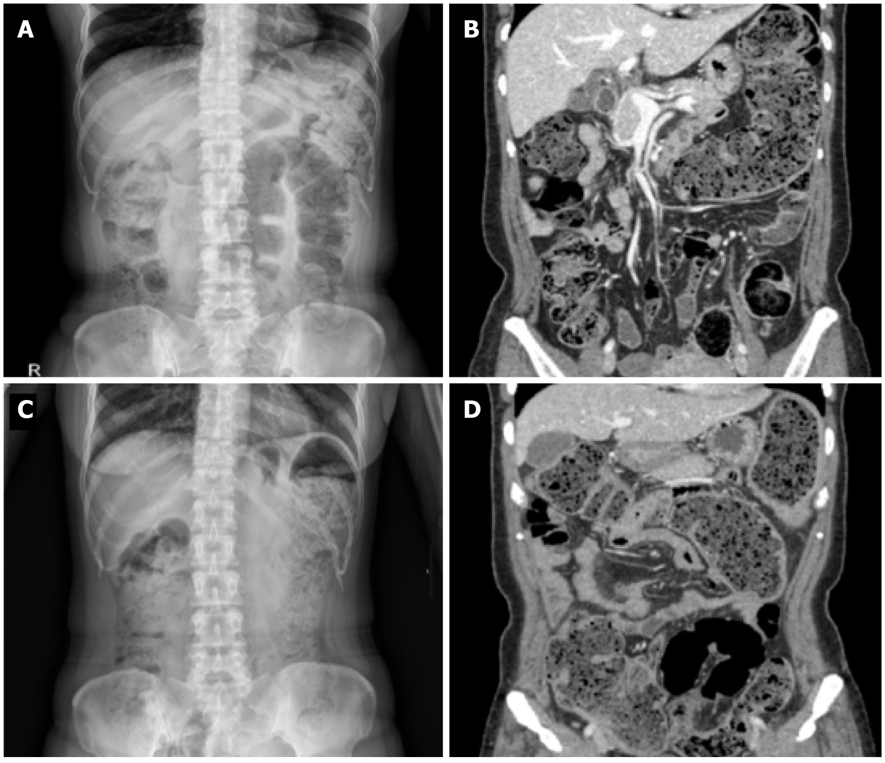
Figure 1 Abdominal X-ray and computed tomography images.
A and B: Abdominal X-ray and computed tomography (CT) scan at initial admission showed fecal impaction and diffuse dilatation of the entire colon; C and D: Abdominal X-ray and CT scan 8 mo after initial admission showed aggravation of fecal impaction and bowel dilatation involving both the ascending and the descending colon with edematous colonic wall thickening in the sigmoid colon.
- Citation: Kim BS, Park SY, Kim DH, Kim NI, Yoon JH, Ju JK, Park CH, Kim HS, Choi SK. Cytomegalovirus colitis induced segmental colonic hypoganglionosis in an immunocompetent patient: A case report. World J Clin Cases 2021; 9(20): 5631-5636
- URL: https://www.wjgnet.com/2307-8960/full/v9/i20/5631.htm
- DOI: https://dx.doi.org/10.12998/wjcc.v9.i20.5631