Copyright
©The Author(s) 2021.
World J Clin Cases. Jul 16, 2021; 9(20): 5621-5630
Published online Jul 16, 2021. doi: 10.12998/wjcc.v9.i20.5621
Published online Jul 16, 2021. doi: 10.12998/wjcc.v9.i20.5621
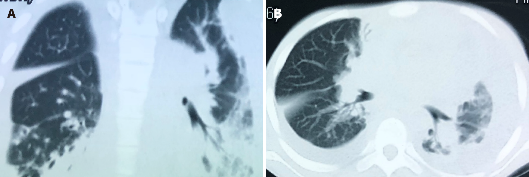
Figure 2 The chest computed tomography on September 21, 2018 shows a massive pleural effusion on the left, a small pleural effusion on the right, patchy and streaky cloudy opacities on both lungs, suggesting inflammation.
A: The sagittal plane; B: The horizontal plane.
- Citation: Liu J, Lei ZY, Pang YH, Huang YX, Xu LJ, Zhu JY, Zheng JX, Yang XH, Lin BL, Gao ZL, Zhuo C. Rapid diagnosis of disseminated Mycobacterium mucogenicum infection in formalin-fixed, paraffin-embedded specimen using next-generation sequencing: A case report. World J Clin Cases 2021; 9(20): 5621-5630
- URL: https://www.wjgnet.com/2307-8960/full/v9/i20/5621.htm
- DOI: https://dx.doi.org/10.12998/wjcc.v9.i20.5621


