
















©The Author(s) 2021.
World J Clin Cases. Jul 16, 2021; 9(20): 5594-5604
Published online Jul 16, 2021. doi: 10.12998/wjcc.v9.i20.5594
Published online Jul 16, 2021. doi: 10.12998/wjcc.v9.i20.5594
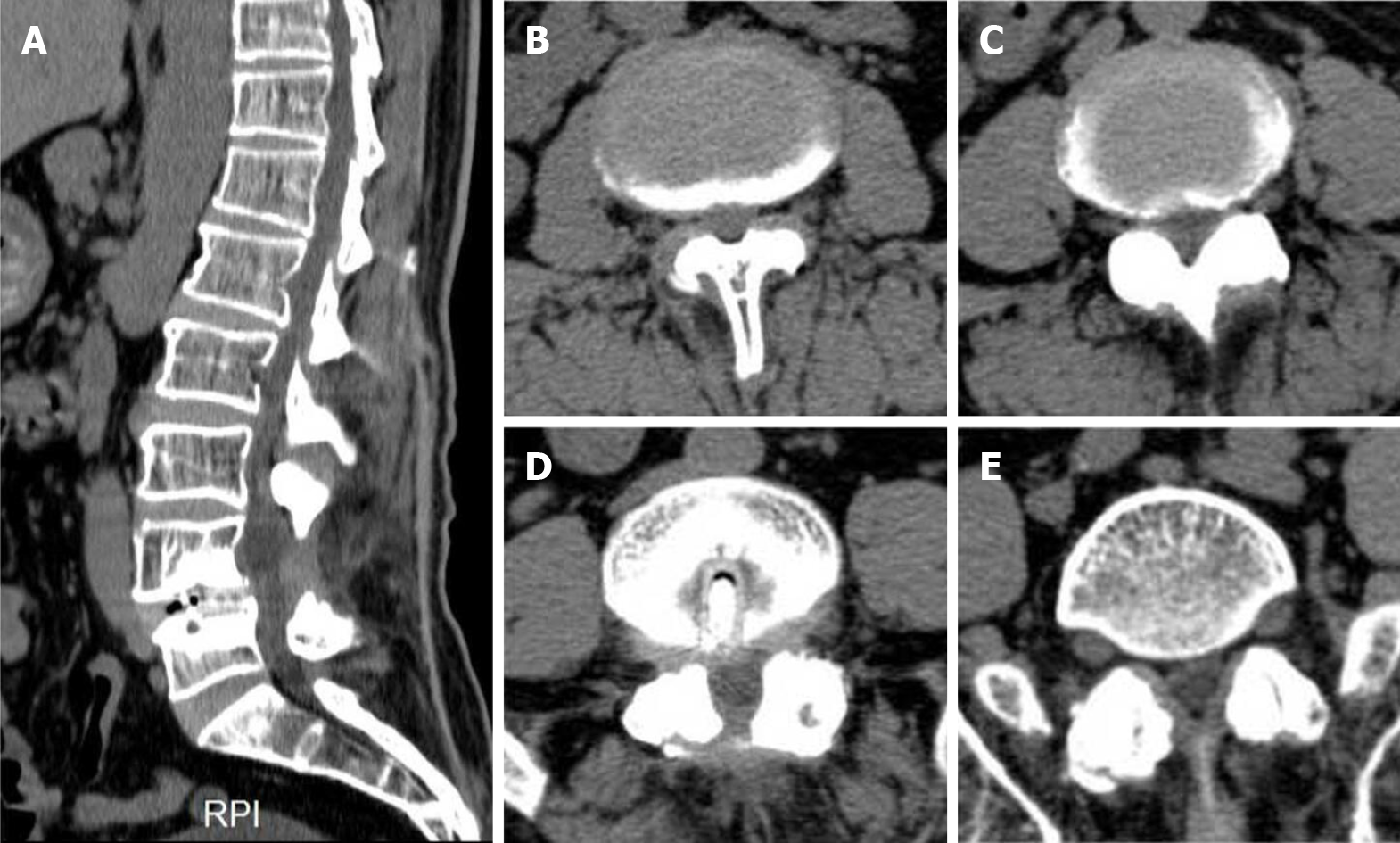
Figure 2 Preoperative computed tomography on the patient’s second admission.
A: Sagittal examination revealed the presence of physiological curvature of the lumbar spine, reduced bone mineral density of all vertebral bodies, irregular margins in some and absence of spinous processes in L4; B-E: L2-L3 (B), L3-L4 (C), L4-L5 (D), L5-S1 (E) cross sections, respectively. Soft tissue density shadowing of multisegmented discs projected towards the periphery. Metal-like structures were found in L4-L5. RPI: Right posterior image.
- Citation: Ouyang Y, Qu Y, Dong RP, Kang MY, Yu T, Cheng XL, Zhao JW. Spinal dural arteriovenous fistula 8 years after lumbar discectomy surgery: A case report and review of literature. World J Clin Cases 2021; 9(20): 5594-5604
- URL: https://www.wjgnet.com/2307-8960/full/v9/i20/5594.htm
- DOI: https://dx.doi.org/10.12998/wjcc.v9.i20.5594