Copyright
©The Author(s) 2021.
World J Clin Cases. Jul 16, 2021; 9(20): 5575-5587
Published online Jul 16, 2021. doi: 10.12998/wjcc.v9.i20.5575
Published online Jul 16, 2021. doi: 10.12998/wjcc.v9.i20.5575
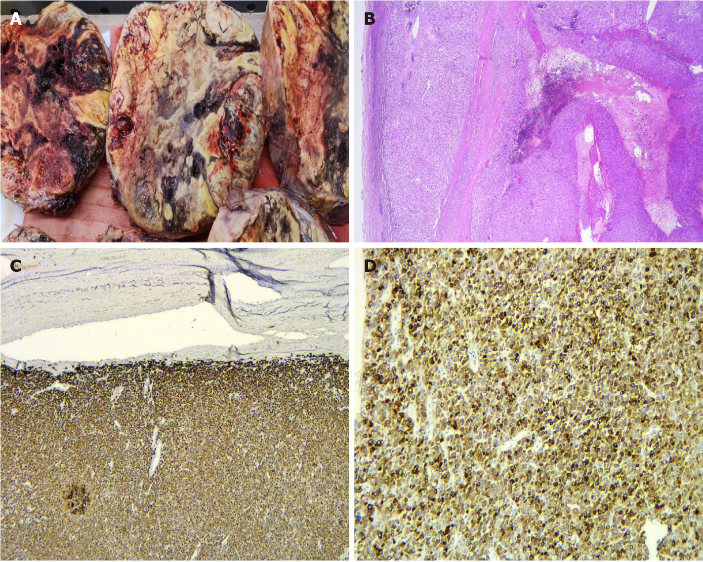
Figure 5 Adrenocortical carcinoma.
A: Large solitary circumscribed tumor with a variegated appearance on the cut surface due to hemorrhage and necrosis; B: Diffuse architecture of the tumor and capsular invasion (hematoxylin & eosin, × 25); C: Intense positivity for Melan A in tumor cells (immunohistochemistry, × 25); D: Intense positivity for synaptophysin (immunohistochemistry, × 200).
- Citation: Costache MF, Arhirii RE, Mogos SJ, Lupascu-Ursulescu C, Litcanu CI, Ciumanghel AI, Cucu C, Ghiciuc CM, Petris AO, Danila N. Giant androgen-producing adrenocortical carcinoma with atrial flutter: A case report and review of the literature. World J Clin Cases 2021; 9(20): 5575-5587
- URL: https://www.wjgnet.com/2307-8960/full/v9/i20/5575.htm
- DOI: https://dx.doi.org/10.12998/wjcc.v9.i20.5575


