Copyright
©The Author(s) 2021.
World J Clin Cases. Jul 16, 2021; 9(20): 5556-5561
Published online Jul 16, 2021. doi: 10.12998/wjcc.v9.i20.5556
Published online Jul 16, 2021. doi: 10.12998/wjcc.v9.i20.5556
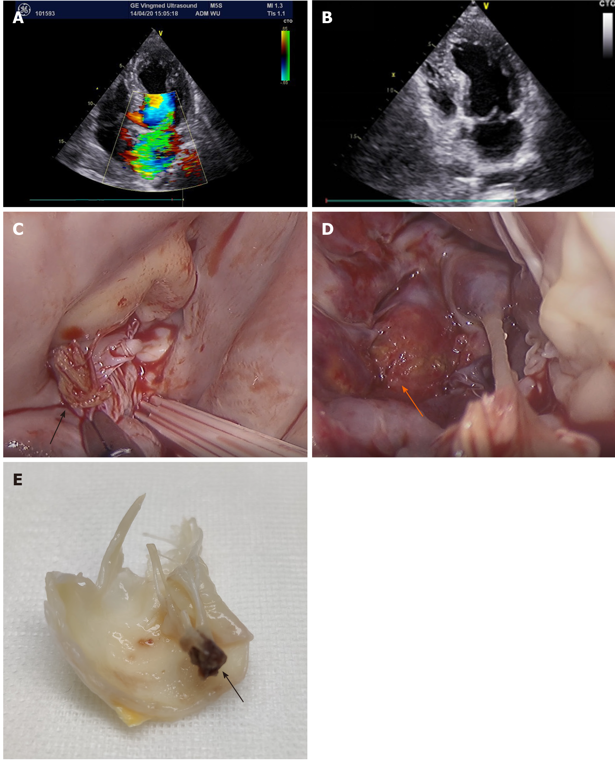
Figure 2 Mitral valve and papillary muscle 2 wk after radiofrequency catheter ablation.
A and B: Echocardiographic image showing massive mitral regurgitation and ruptured mitral valve leaflet which was entering into the left atrium; C and D: The rupture of papillary muscles was found during mitral valve replacement; E: The mitral valve was cut off during the surgery. Arrows show the end of the rupture of papillary muscles.
- Citation: Sun ZW, Wu BF, Ying X, Zhang BQ, Yao L, Zheng LR. Delayed papillary muscle rupture after radiofrequency catheter ablation: A case report. World J Clin Cases 2021; 9(20): 5556-5561
- URL: https://www.wjgnet.com/2307-8960/full/v9/i20/5556.htm
- DOI: https://dx.doi.org/10.12998/wjcc.v9.i20.5556


