Copyright
©The Author(s) 2021.
World J Clin Cases. Jul 16, 2021; 9(20): 5462-5469
Published online Jul 16, 2021. doi: 10.12998/wjcc.v9.i20.5462
Published online Jul 16, 2021. doi: 10.12998/wjcc.v9.i20.5462
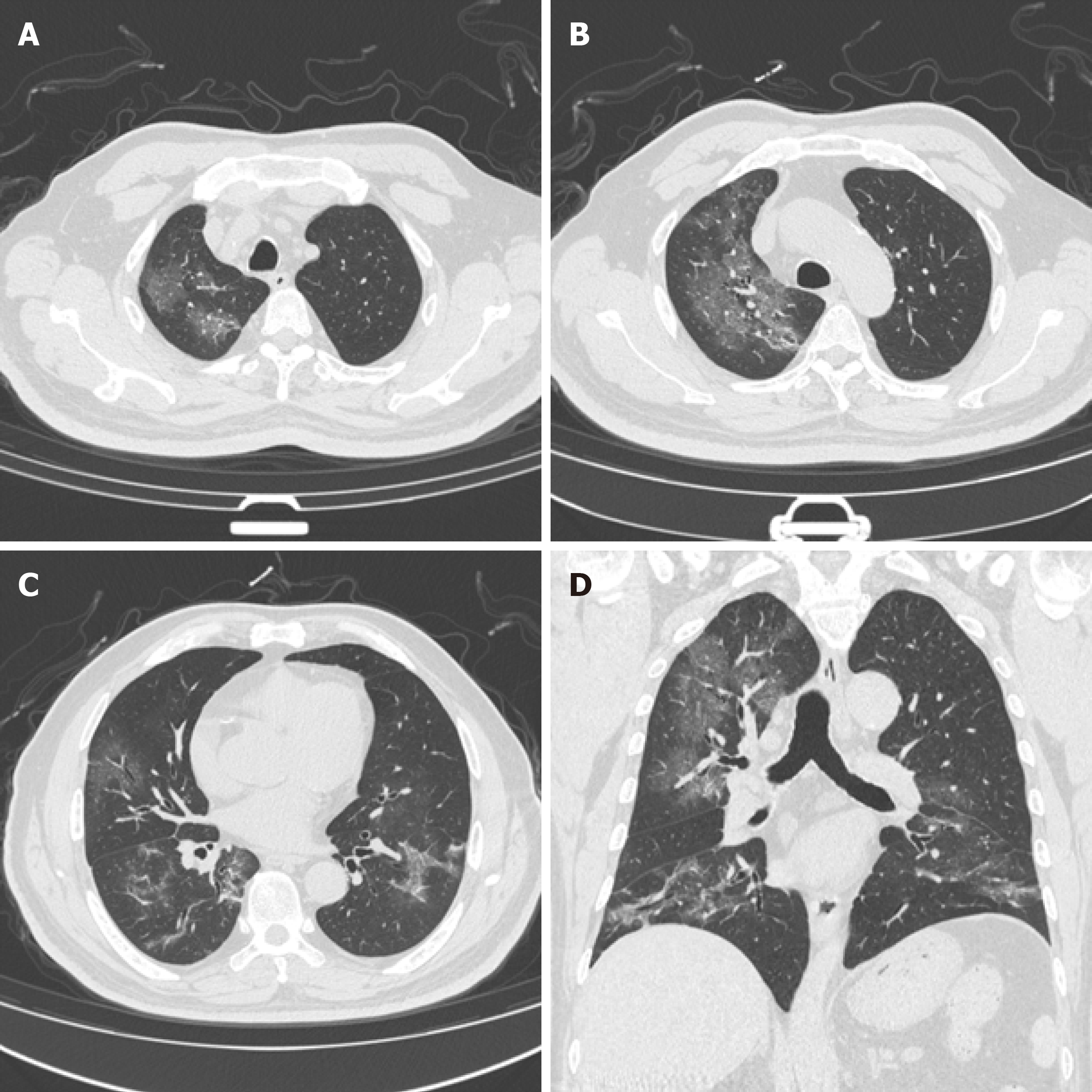
Figure 1 CT images from a 61-year-old male patient with a total lung score of 16 obtained 14 d after the onset of disease.
A: A large ground glass density shadow of the right upper lobe; B: A large ground glass density of the right upper lobe with a small amount of flaky consolidation; C: A ground glass density shadow of the middle lobe of the right lung, paving-stone lesions of the lower lobe of the left lung, and a striped shadow in the lower lobe of both lungs; D: Coronal reconstruction images show a ground glass density shadow in the right upper lung. Both lower lungs are dominated by fibrous strips.
- Citation: Li XL, Li T, Du QC, Yang L, He KL. Effects of angiotensin receptor blockers and angiotensin-converting enzyme inhibitors on COVID-19. World J Clin Cases 2021; 9(20): 5462-5469
- URL: https://www.wjgnet.com/2307-8960/full/v9/i20/5462.htm
- DOI: https://dx.doi.org/10.12998/wjcc.v9.i20.5462


