Copyright
©The Author(s) 2021.
World J Clin Cases. Jan 16, 2021; 9(2): 516-520
Published online Jan 16, 2021. doi: 10.12998/wjcc.v9.i2.516
Published online Jan 16, 2021. doi: 10.12998/wjcc.v9.i2.516
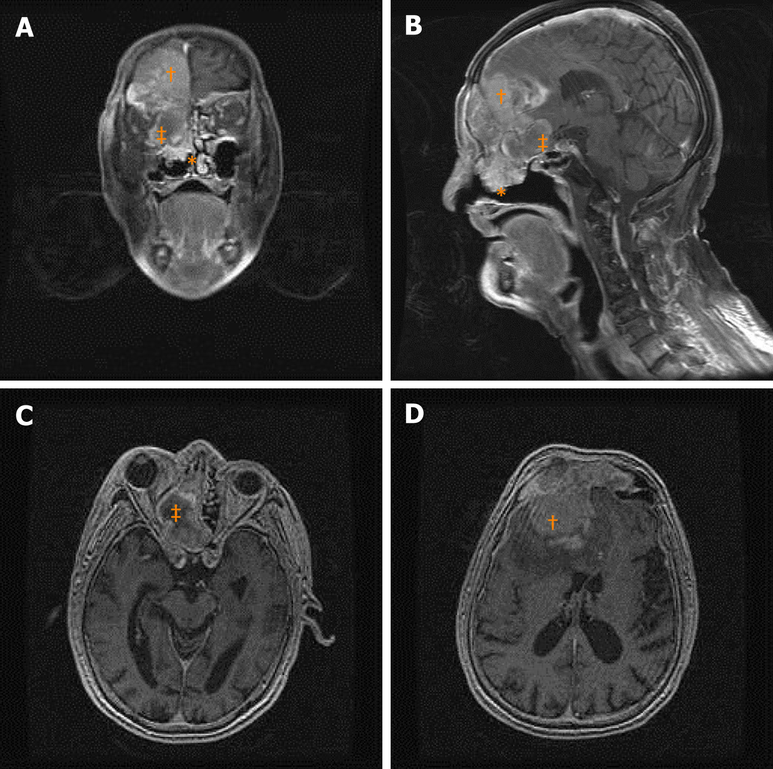
Figure 1 Magnetic resonance imaging of the head with contrast showed a heterogeneously enhanced mass involving the right nasal cavity (asterisk), orbital cavity, and bilateral frontal sinus with intracranial invasion (cross), and another lobulated fluid collection, suspected as a mucocele, at the right ethmoid cells (double-cross).
A: Coronal section; B: Sagittal section; C: Axial section of the magnetic resonance imaging, which showed the orbital cavity invaded and compressed by the lesion (double-cross) from the medial side; D: Bilateral frontal sinuses that were involved by the lesion.
- Citation: Hsu HJ, Huang CC, Chuang MT, Tien CH, Lee JS, Lee PH. Recurrent inverted papilloma coexisted with skull base lymphoma: A case report. World J Clin Cases 2021; 9(2): 516-520
- URL: https://www.wjgnet.com/2307-8960/full/v9/i2/516.htm
- DOI: https://dx.doi.org/10.12998/wjcc.v9.i2.516


