
















©The Author(s) 2021.
World J Clin Cases. Jan 16, 2021; 9(2): 509-515
Published online Jan 16, 2021. doi: 10.12998/wjcc.v9.i2.509
Published online Jan 16, 2021. doi: 10.12998/wjcc.v9.i2.509
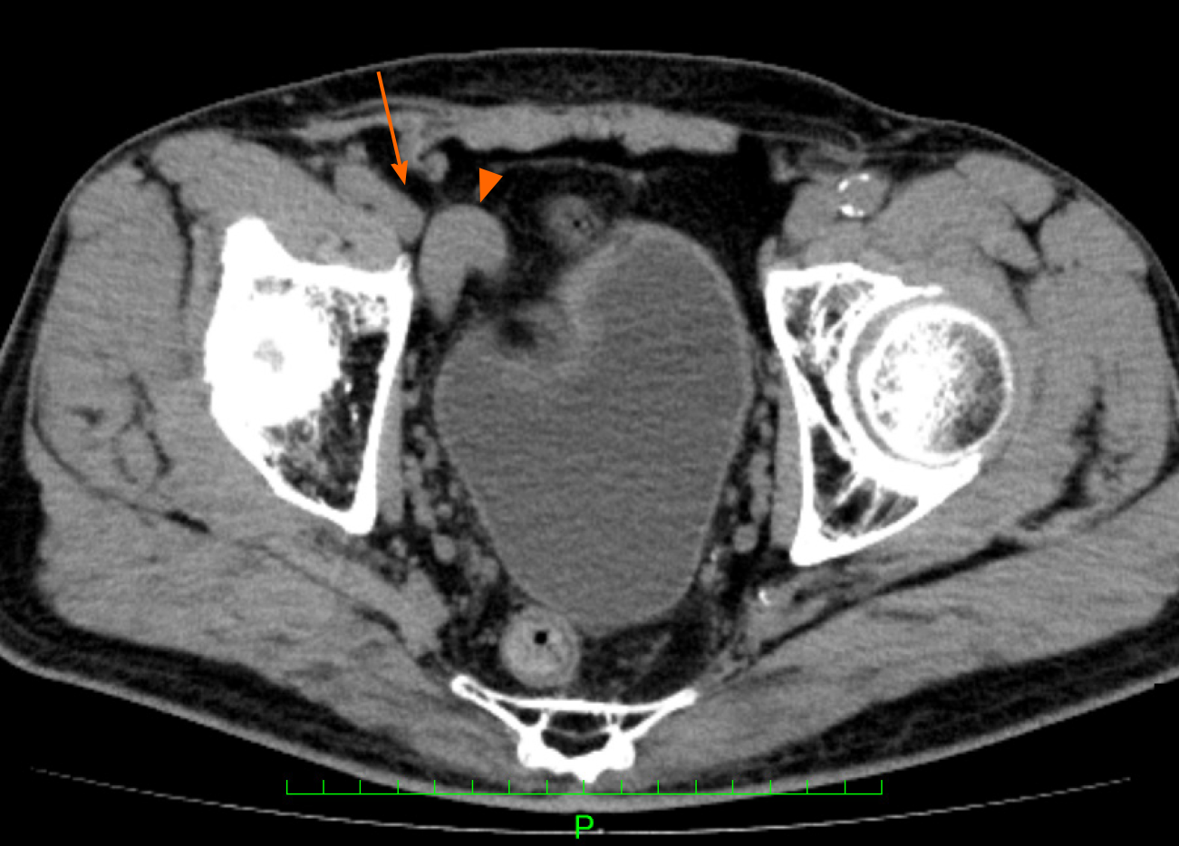
Figure 4 Postoperative computed tomography findings.
The computed tomography scans indicated that the shunt vessel was no longer located near the right internal inguinal ring, and it had separated from the femoral vein. Triangle: Shunt vessel; Arrow: Femoral vein.
- Citation: Yura M, Yo K, Hara A, Hayashi K, Tajima Y, Kaneko Y, Fujisaki H, Hirata A, Takano K, Hongo K, Yoneyama K, Nakagawa M. Indirect inguinal hernia containing portosystemic shunt vessel: A case report. World J Clin Cases 2021; 9(2): 509-515
- URL: https://www.wjgnet.com/2307-8960/full/v9/i2/509.htm
- DOI: https://dx.doi.org/10.12998/wjcc.v9.i2.509