Copyright
©The Author(s) 2021.
World J Clin Cases. Jan 16, 2021; 9(2): 509-515
Published online Jan 16, 2021. doi: 10.12998/wjcc.v9.i2.509
Published online Jan 16, 2021. doi: 10.12998/wjcc.v9.i2.509
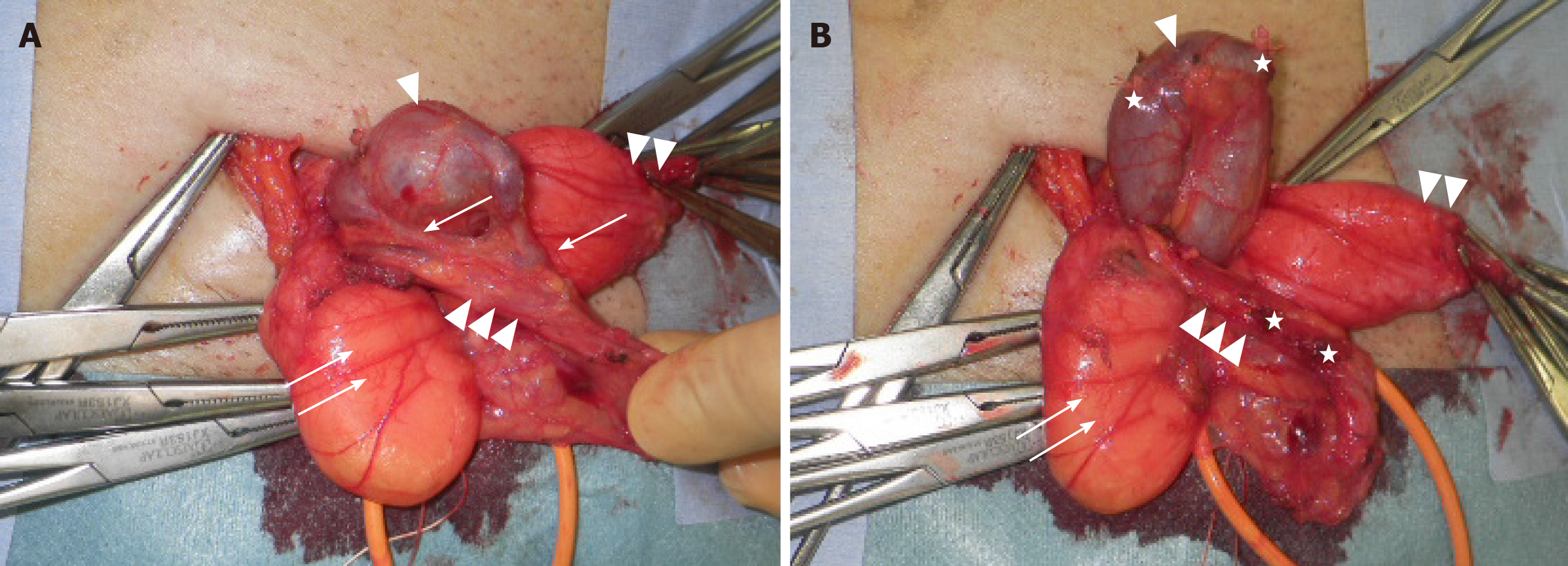
Figure 3 Photograph taken during surgery.
A: The expanded (2-cm-diameter) shunt vessel existing in the extraperitoneal cavity entered the inguinal canal through the internal inguinal ring. An indirect hernia sac and two short branches connecting the shunt vessel and the testicular vein were identified; B: Shunt vessel after cutting short branches. Triangle: Shunt vessel; Double triangles: Indirect hernia sac; Triple triangles: Vas deferens and testicular artery and vein; Arrow: Two short branches between the shunt vessel and testicular vein; Double arrows: Extraperitoneal fat; Stars: Cut-off stumps of ligated short branches.
- Citation: Yura M, Yo K, Hara A, Hayashi K, Tajima Y, Kaneko Y, Fujisaki H, Hirata A, Takano K, Hongo K, Yoneyama K, Nakagawa M. Indirect inguinal hernia containing portosystemic shunt vessel: A case report. World J Clin Cases 2021; 9(2): 509-515
- URL: https://www.wjgnet.com/2307-8960/full/v9/i2/509.htm
- DOI: https://dx.doi.org/10.12998/wjcc.v9.i2.509


