Copyright
©The Author(s) 2021.
World J Clin Cases. Jan 16, 2021; 9(2): 463-468
Published online Jan 16, 2021. doi: 10.12998/wjcc.v9.i2.463
Published online Jan 16, 2021. doi: 10.12998/wjcc.v9.i2.463
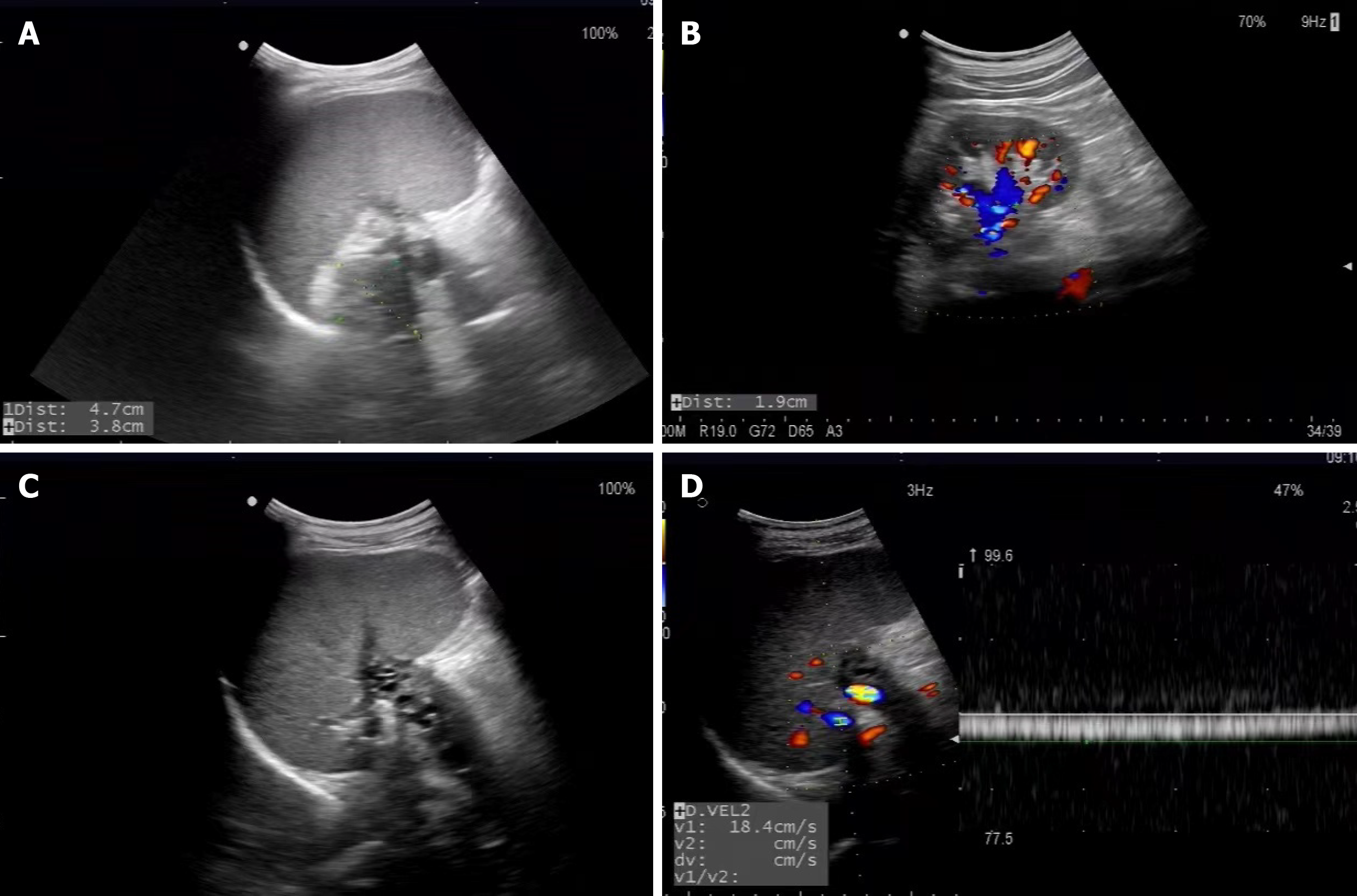
Figure 1 Ultrasonography imaging.
A: Pancreatic pseudocysts with the cystic heterogeneous hypoechoic area; B: Renal venous congestion; C: The formation of splenic venous collateral and main splenic venous thrombus; D: The peak velocity of splenic venous collateral with continuous waveform.
- Citation: Chen BB, Mu PY, Lu JT, Wang G, Zhang R, Huang DD, Shen DH, Jiang TT. Sinistral portal hypertension associated with pancreatic pseudocysts - ultrasonography findings: A case report. World J Clin Cases 2021; 9(2): 463-468
- URL: https://www.wjgnet.com/2307-8960/full/v9/i2/463.htm
- DOI: https://dx.doi.org/10.12998/wjcc.v9.i2.463


