Copyright
©The Author(s) 2021.
World J Clin Cases. Jan 16, 2021; 9(2): 416-421
Published online Jan 16, 2021. doi: 10.12998/wjcc.v9.i2.416
Published online Jan 16, 2021. doi: 10.12998/wjcc.v9.i2.416
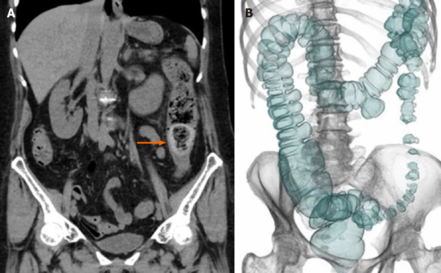
Figure 2 Computed tomography.
A: The coronal image shows a fecalith impaction at the sigmoid-descending junction (arrow); B: Computed tomography colonography shows a large diverticulum in the transverse colon and mild stenosis with thumb printing in the descending colon.
- Citation: Tanabe H, Tanaka K, Goto M, Sato T, Sato K, Fujiya M, Okumura T. Rare case of fecal impaction caused by a fecalith originating in a large colonic diverticulum: A case report. World J Clin Cases 2021; 9(2): 416-421
- URL: https://www.wjgnet.com/2307-8960/full/v9/i2/416.htm
- DOI: https://dx.doi.org/10.12998/wjcc.v9.i2.416


