Copyright
©The Author(s) 2021.
World J Clin Cases. Jan 16, 2021; 9(2): 403-409
Published online Jan 16, 2021. doi: 10.12998/wjcc.v9.i2.403
Published online Jan 16, 2021. doi: 10.12998/wjcc.v9.i2.403
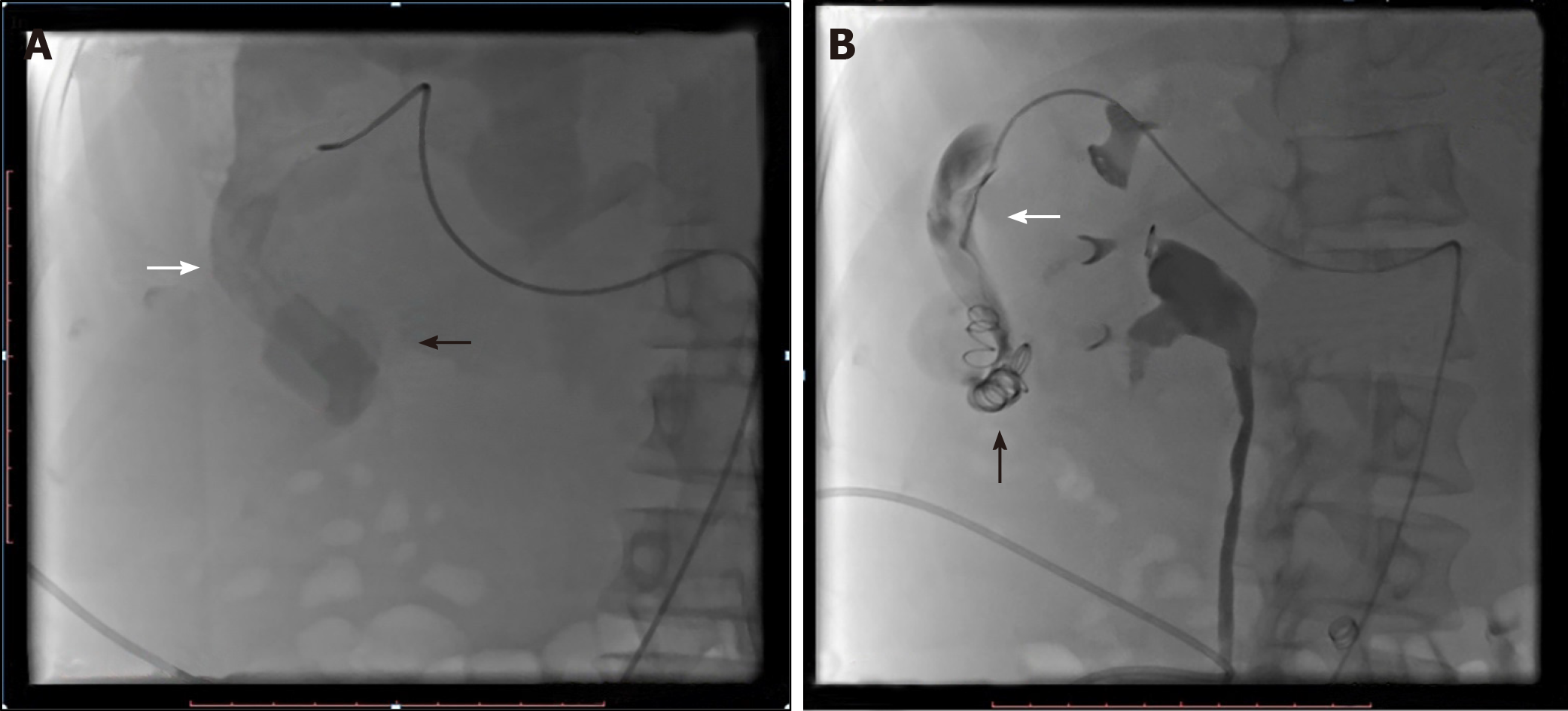
Figure 2 Selective right hepatic angiogram before and after coil embolization.
A: Angiography demonstrated the hypertrophied right hepatic artery (white arrow) and vascular malformation (black arrow) around the arterioportal fistula. B: Angiography showed the catheter in the enlarged right hepatic artery (white arrow). The distal segments of the right hepatic artery were occluded using two 12-3 and six 8-5 coils (black arrow).
- Citation: Stepanyan SA, Poghosyan T, Manukyan K, Hakobyan G, Hovhannisyan H, Safaryan H, Baghdasaryan E, Gemilyan M. Coil embolization of arterioportal fistula complicated by gastrointestinal bleeding after Caesarian section: A case report. World J Clin Cases 2021; 9(2): 403-409
- URL: https://www.wjgnet.com/2307-8960/full/v9/i2/403.htm
- DOI: https://dx.doi.org/10.12998/wjcc.v9.i2.403


