Copyright
©The Author(s) 2021.
World J Clin Cases. Jan 16, 2021; 9(2): 396-402
Published online Jan 16, 2021. doi: 10.12998/wjcc.v9.i2.396
Published online Jan 16, 2021. doi: 10.12998/wjcc.v9.i2.396
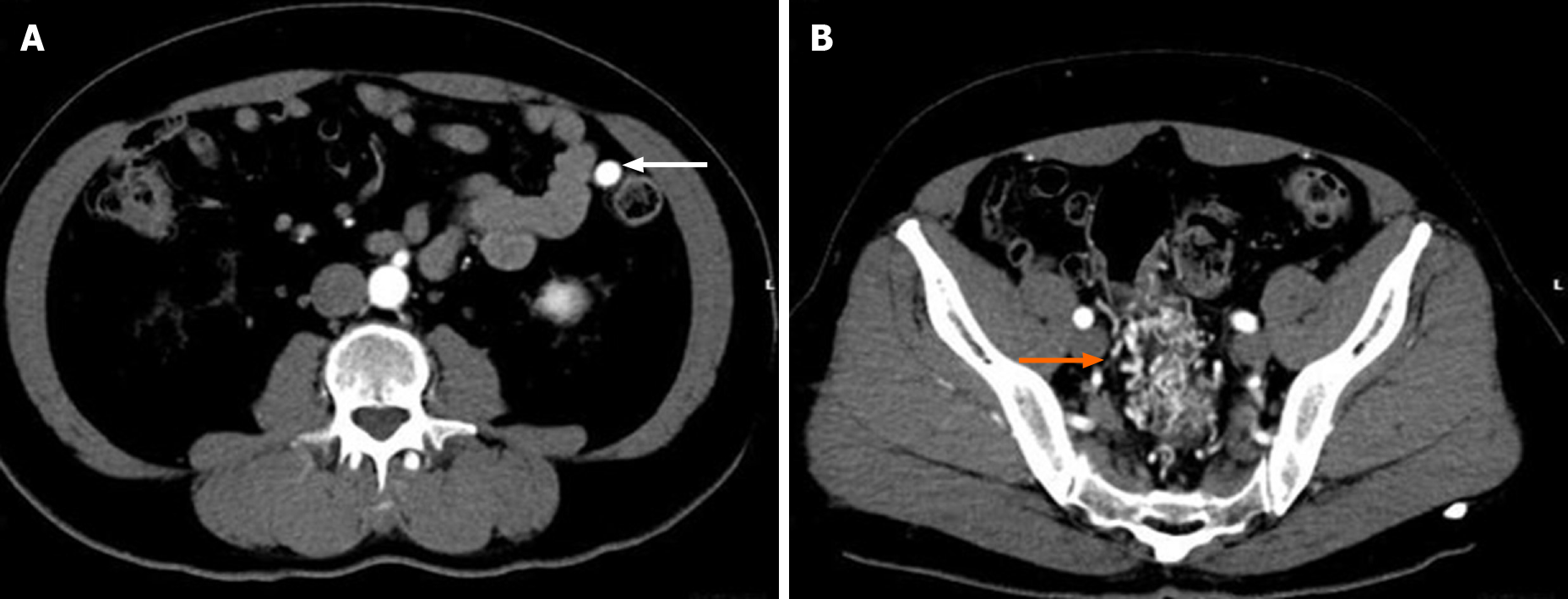
Figure 2 Images from contrast-enhanced abdominal computed tomography.
A contrast-enhanced abdominal computed tomography scan showed the nidus of small corkscrew and dilated vessels (orange arrow) and marked early inferior mesenteric vein enhancement (white arrow).
- Citation: Kimura Y, Hara T, Nagao R, Nakanishi T, Kawaguchi J, Tagami A, Ikeda T, Araki H, Tsurumi H. Natural history of inferior mesenteric arteriovenous malformation that led to ischemic colitis: A case report. World J Clin Cases 2021; 9(2): 396-402
- URL: https://www.wjgnet.com/2307-8960/full/v9/i2/396.htm
- DOI: https://dx.doi.org/10.12998/wjcc.v9.i2.396


