
















©The Author(s) 2021.
World J Clin Cases. Jul 6, 2021; 9(19): 5332-5338
Published online Jul 6, 2021. doi: 10.12998/wjcc.v9.i19.5332
Published online Jul 6, 2021. doi: 10.12998/wjcc.v9.i19.5332
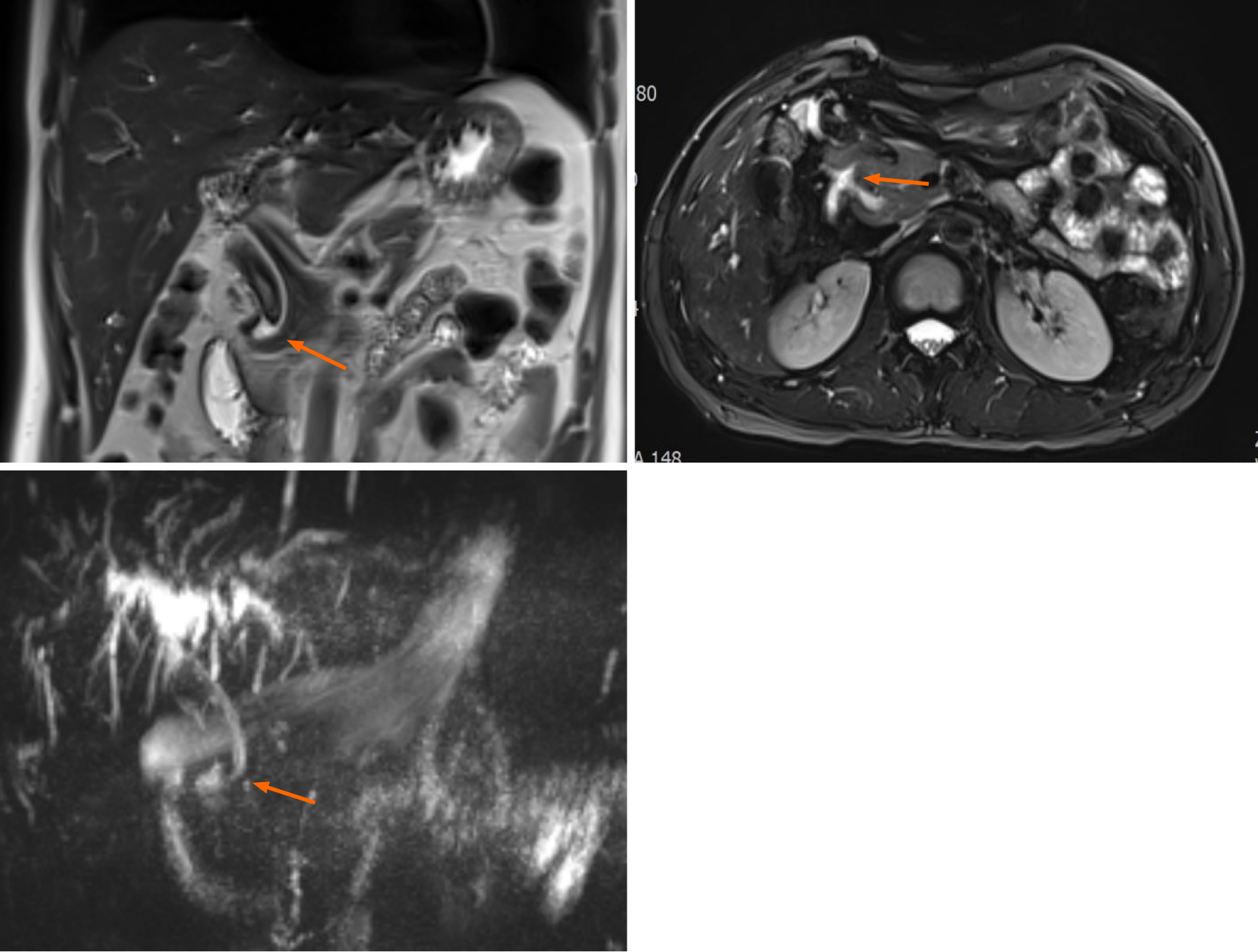
Figure 2 Magnetic resonance imaging findings.
Magnetic resonance imaging showed a hook-shaped change at the lower end of the common bile duct, which merged into the lower posterior wall of the duodenal bulb. The orange arrow shows the hook shape.
- Citation: Xu H, Li X, Zhu KX, Zhou WC. Ectopic opening of the common bile duct into the duodenal bulb with recurrent choledocholithiasis: A case report. World J Clin Cases 2021; 9(19): 5332-5338
- URL: https://www.wjgnet.com/2307-8960/full/v9/i19/5332.htm
- DOI: https://dx.doi.org/10.12998/wjcc.v9.i19.5332