Copyright
©The Author(s) 2021.
World J Clin Cases. Jul 6, 2021; 9(19): 5259-5265
Published online Jul 6, 2021. doi: 10.12998/wjcc.v9.i19.5259
Published online Jul 6, 2021. doi: 10.12998/wjcc.v9.i19.5259
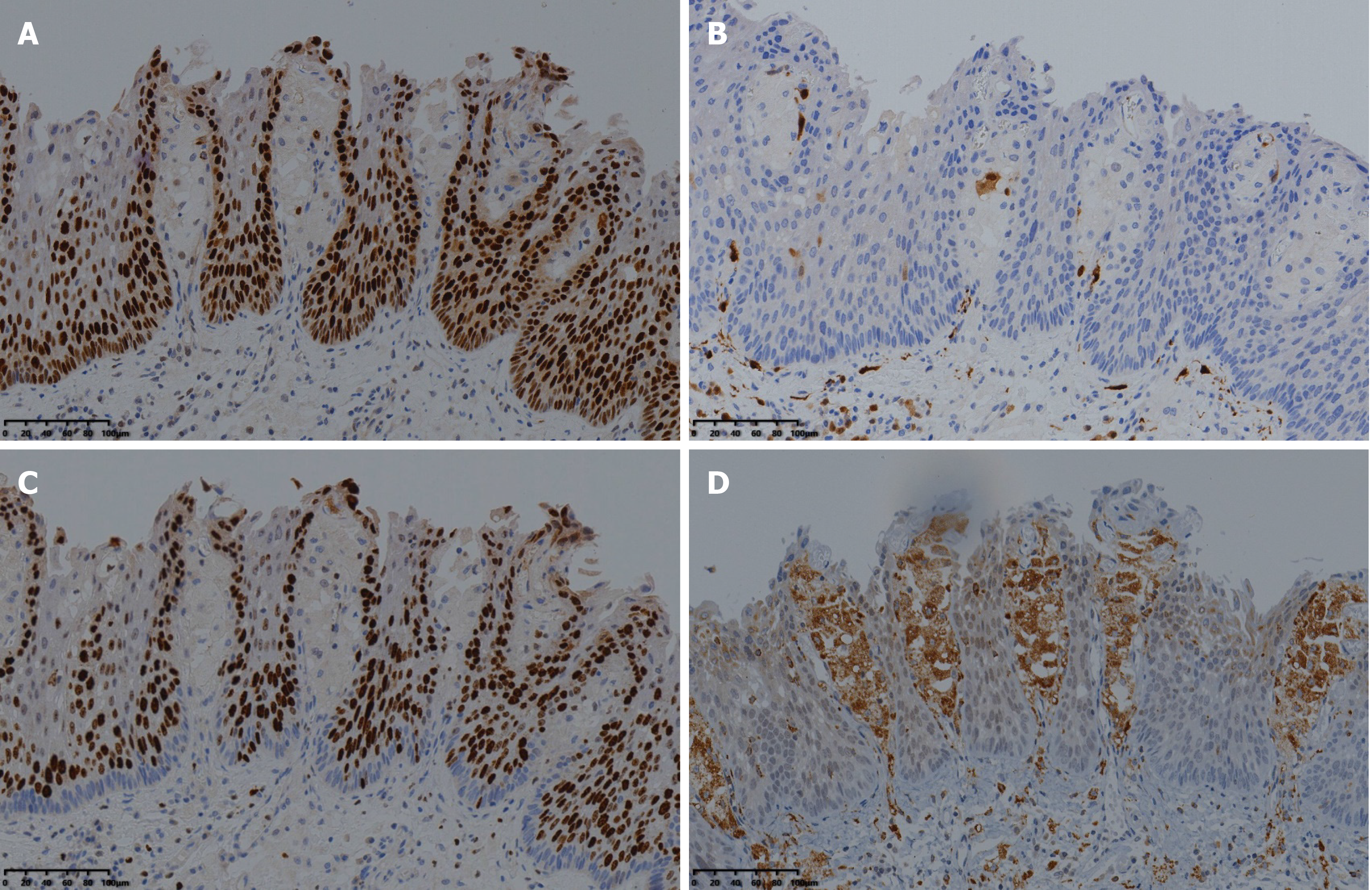
Figure 4 Immunohistochemical findings.
A and B: Immunohistochemical staining showed that the regions of squamous cell carcinoma were positive for P53 and negative for P16 (× 20); C: The Ki-67 index was 90% (× 20); D: The observed foam cells were strongly positive for CD68 (× 20).
- Citation: Yang XY, Fu KI, Chen YP, Chen ZW, Ding J. Diffuse xanthoma in early esophageal cancer: A case report. World J Clin Cases 2021; 9(19): 5259-5265
- URL: https://www.wjgnet.com/2307-8960/full/v9/i19/5259.htm
- DOI: https://dx.doi.org/10.12998/wjcc.v9.i19.5259


