
















©The Author(s) 2021.
World J Clin Cases. Jul 6, 2021; 9(19): 5211-5216
Published online Jul 6, 2021. doi: 10.12998/wjcc.v9.i19.5211
Published online Jul 6, 2021. doi: 10.12998/wjcc.v9.i19.5211
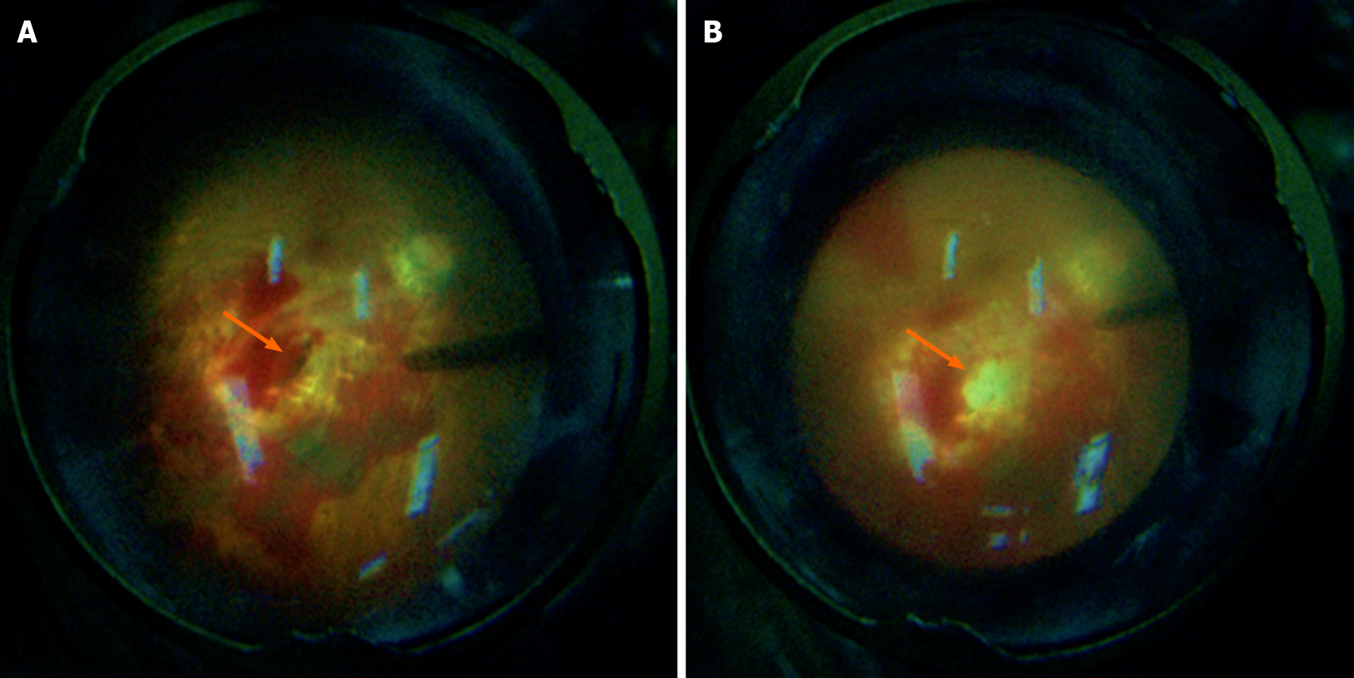
Figure 3 Intraoperative fundus photographs.
A: Intraoperative fundus photograph showing a penetrating port with a transverse diameter of about one pupillary distance (PD) seen in the eyeball wall at two PD below the temporal region of the macula in the right eye; B: The autologous fascia was packed in the posterior exit wound, on which the surrounding inner limiting membrane was peeled off, flipped, and covered during the operation.
- Citation: Yi QY, Wang SS, Gui Q, Chen LS, Li WD. Autologous tenon capsule packing to treat posterior exit wound of penetrating injury: A case report. World J Clin Cases 2021; 9(19): 5211-5216
- URL: https://www.wjgnet.com/2307-8960/full/v9/i19/5211.htm
- DOI: https://dx.doi.org/10.12998/wjcc.v9.i19.5211