Copyright
©The Author(s) 2021.
World J Clin Cases. Jun 26, 2021; 9(18): 4772-4777
Published online Jun 26, 2021. doi: 10.12998/wjcc.v9.i18.4772
Published online Jun 26, 2021. doi: 10.12998/wjcc.v9.i18.4772
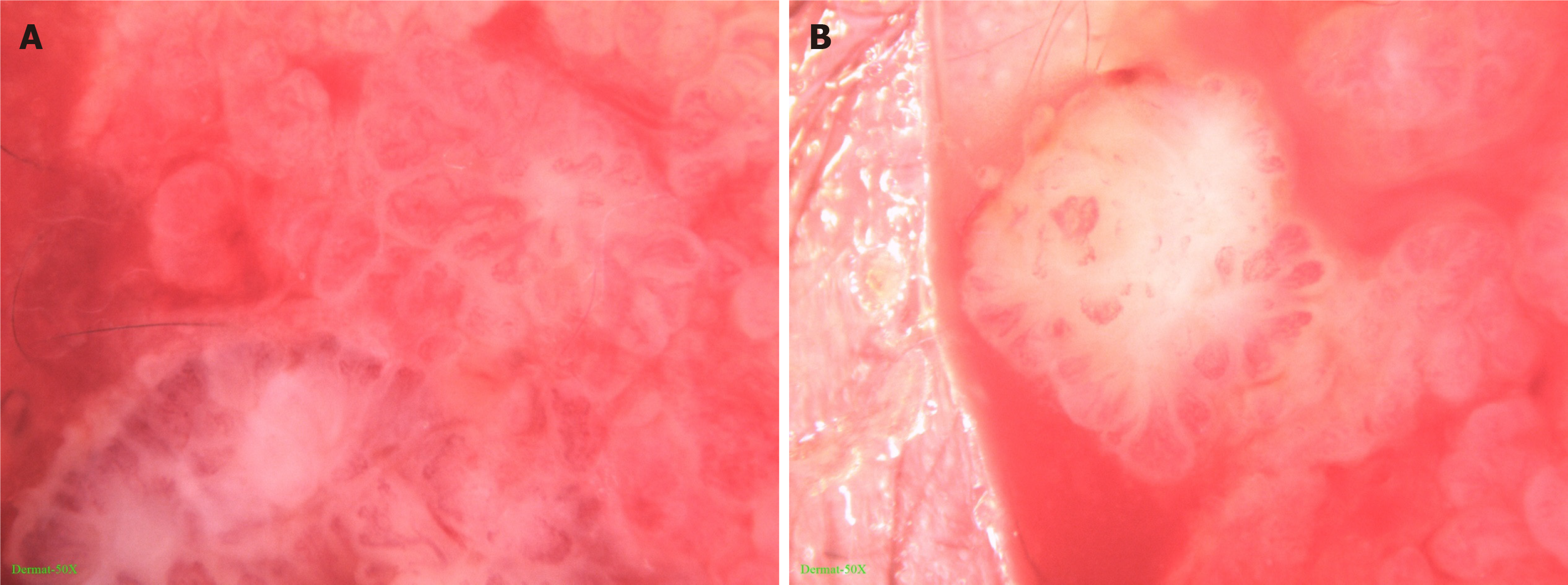
Figure 2 Dermatoscopic appearance of the lesion.
The infiltration method was used (× 50). A: The skin lesion had lobular and crumby structures. Its center was reddish or white while the edges were white or yellowish, band-like; B: There were polymorphic vascular structures and white radial streaks in the lesion, with some vascular clusters scattered.
- Citation: Jiang HJ, Zhang Z, Zhang L, Pu YJ, Zhou N, Shu H. Neonatal syringocystadenoma papilliferum: A case report. World J Clin Cases 2021; 9(18): 4772-4777
- URL: https://www.wjgnet.com/2307-8960/full/v9/i18/4772.htm
- DOI: https://dx.doi.org/10.12998/wjcc.v9.i18.4772


