Copyright
©The Author(s) 2021.
World J Clin Cases. Jun 26, 2021; 9(18): 4741-4747
Published online Jun 26, 2021. doi: 10.12998/wjcc.v9.i18.4741
Published online Jun 26, 2021. doi: 10.12998/wjcc.v9.i18.4741
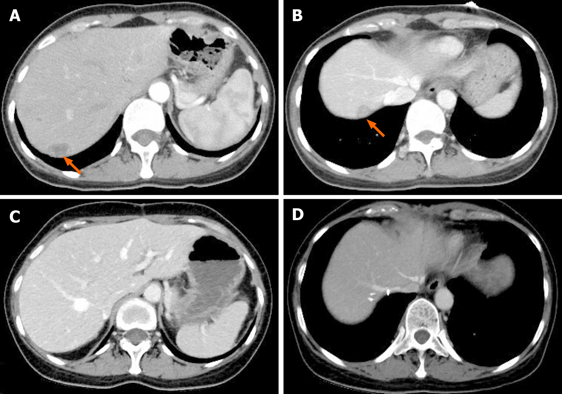
Figure 4 Computed tomography images of the patient with hepatic recurrence.
A and B: Abdominal computed tomography (CT) scan detected low-density nodules under the liver capsule. The arrows indicate metastatic nodules in the liver, and the diameters of the metastatic lesions ranged from 1.5-2.5 cm; C and D: CT appearances of the liver after second resection of tumors in the liver plus partial hepatectomy.
- Citation: Xie C, Shen YM, Chen QH, Bian C. Primary mesonephric adenocarcinoma of the fallopian tube: A case report. World J Clin Cases 2021; 9(18): 4741-4747
- URL: https://www.wjgnet.com/2307-8960/full/v9/i18/4741.htm
- DOI: https://dx.doi.org/10.12998/wjcc.v9.i18.4741


