Copyright
©The Author(s) 2021.
World J Clin Cases. Jun 26, 2021; 9(18): 4741-4747
Published online Jun 26, 2021. doi: 10.12998/wjcc.v9.i18.4741
Published online Jun 26, 2021. doi: 10.12998/wjcc.v9.i18.4741
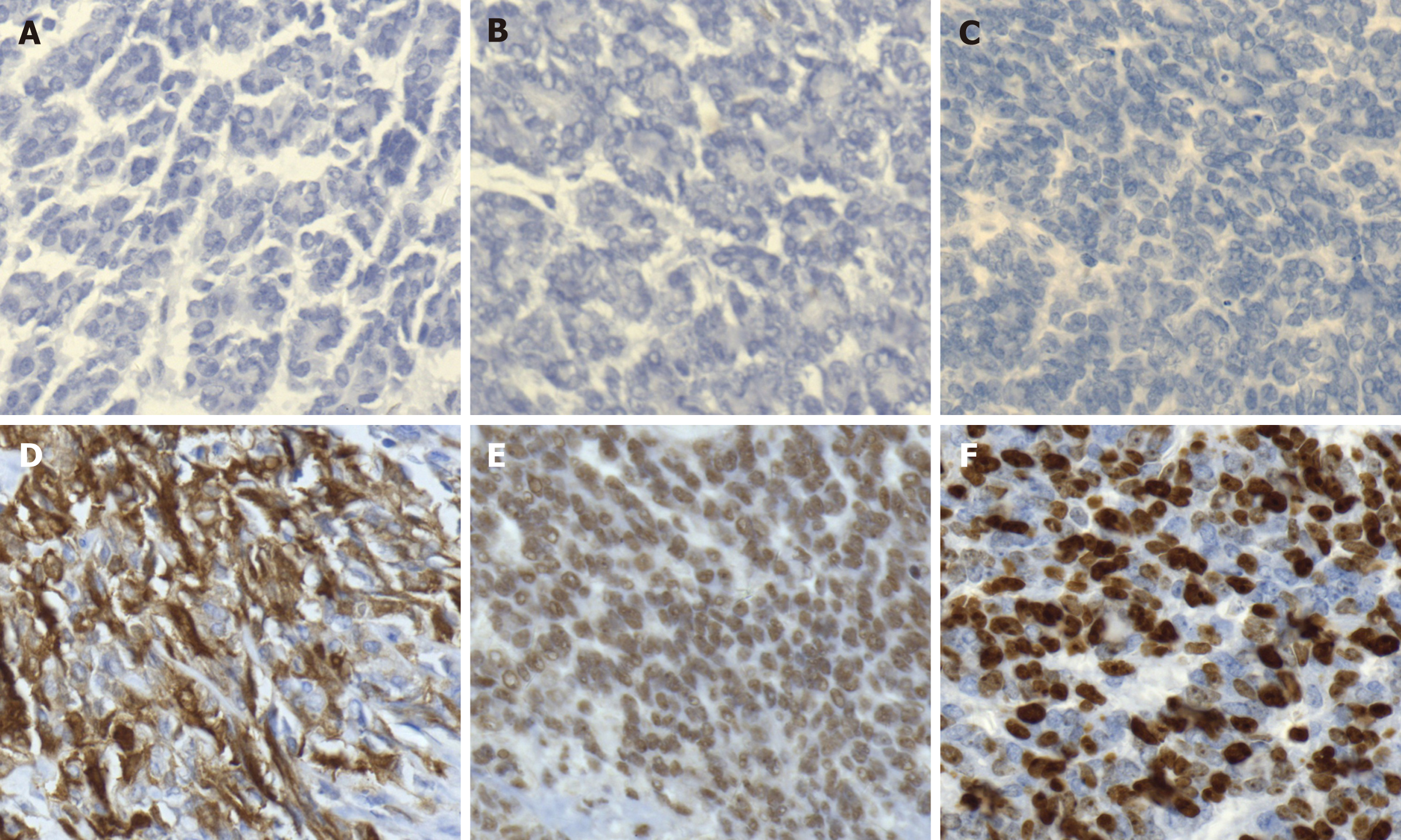
Figure 3 Immunohistochemical staining of fallopian tube-mesonephric adenocarcinoma.
A-C: Immunohistochemistry demonstrated totally negative staining for estrogen receptor (A), progesterone receptor (B), and CA125 (C); D and E: Positive staining for P16 (D) and P53 (E) was detected; F: The positive rate of Ki-67 expression was 60%-70%.
- Citation: Xie C, Shen YM, Chen QH, Bian C. Primary mesonephric adenocarcinoma of the fallopian tube: A case report. World J Clin Cases 2021; 9(18): 4741-4747
- URL: https://www.wjgnet.com/2307-8960/full/v9/i18/4741.htm
- DOI: https://dx.doi.org/10.12998/wjcc.v9.i18.4741


