Copyright
©The Author(s) 2021.
World J Clin Cases. Jun 16, 2021; 9(17): 4433-4440
Published online Jun 16, 2021. doi: 10.12998/wjcc.v9.i17.4433
Published online Jun 16, 2021. doi: 10.12998/wjcc.v9.i17.4433
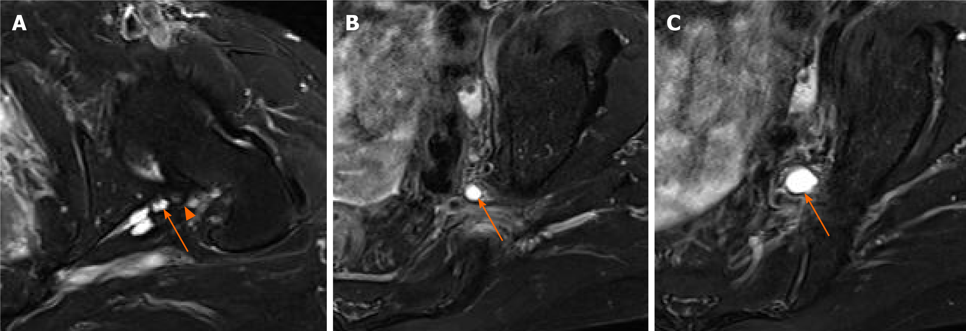
Figure 3 Magnetic resonance imaging.
A: A fat-suppressed T2-weighted axial image showing a degenerative change in the posterior labrum (arrowhead) and the paralabral cyst (arrow); B and C: Rostral to the level of the sciatic notch, the cyst (arrow) was more likely to be expansile and had a balloon-like feature.
- Citation: Lee JG, Peo H, Cho JH, Kim DH. Intraneural ganglion cyst of the lumbosacral plexus mimicking L5 radiculopathy: A case report. World J Clin Cases 2021; 9(17): 4433-4440
- URL: https://www.wjgnet.com/2307-8960/full/v9/i17/4433.htm
- DOI: https://dx.doi.org/10.12998/wjcc.v9.i17.4433


