Copyright
©The Author(s) 2021.
World J Clin Cases. Jun 16, 2021; 9(17): 4365-4372
Published online Jun 16, 2021. doi: 10.12998/wjcc.v9.i17.4365
Published online Jun 16, 2021. doi: 10.12998/wjcc.v9.i17.4365
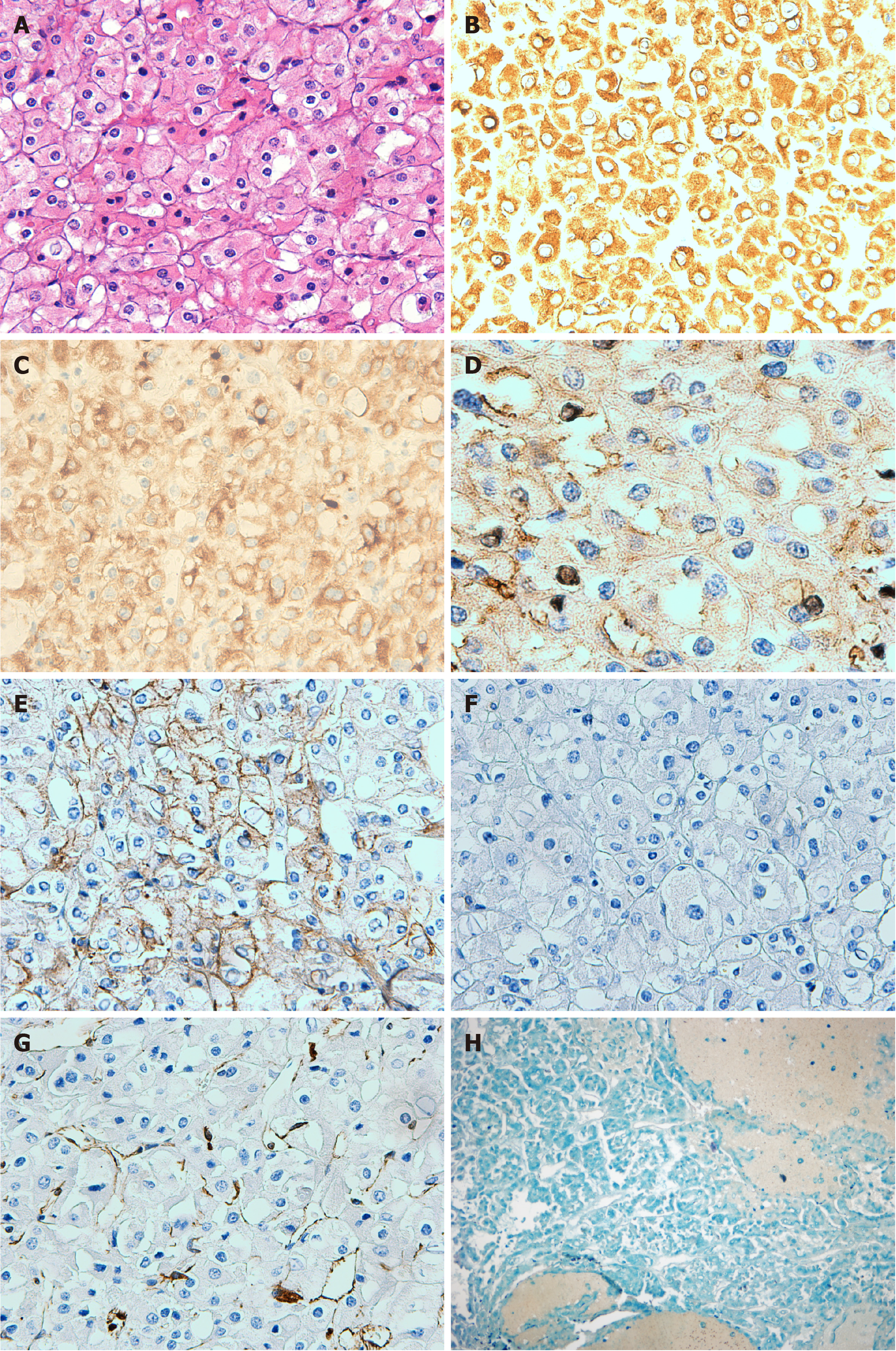
Figure 2 Microscopic photographs of the renal graft tumor.
A: Hematoxylin and eosin staining shows large and polygonal tumor cells, with clearly membrane and polymorphic nuclei. Binucleation can be seen (200 ×); B: Immunohistochemical staining shows positive expression of cytokeratin 7 (200 ×); C: Immunohistochemical staining shows tumor cells positive for cytokeratin 8 (200 ×); D: Immunohistochemical staining shows that cluster of differentiation (CD) 117 is diffusely positive in the cytoplasm/membrane (400 ×); E: Immunohistochemical staining shows that E-cadherin is positive (200 ×); F: Immunohistochemical staining shows that tumor cells are negative for CD10 (200 ×); G: Immunohistochemical staining shows that tumor cells are negative for vimentin (200 ×); H: The cytoplasm of tumor cells appear blue on Hale colloidal iron staining (40 ×).
- Citation: Wang H, Song WL, Cai WJ, Feng G, Fu YX. Kidney re-transplantation after living donor graft nephrectomy due to de novo chromophobe renal cell carcinoma: A case report. World J Clin Cases 2021; 9(17): 4365-4372
- URL: https://www.wjgnet.com/2307-8960/full/v9/i17/4365.htm
- DOI: https://dx.doi.org/10.12998/wjcc.v9.i17.4365


