Copyright
©The Author(s) 2021.
World J Clin Cases. Jun 16, 2021; 9(17): 4327-4335
Published online Jun 16, 2021. doi: 10.12998/wjcc.v9.i17.4327
Published online Jun 16, 2021. doi: 10.12998/wjcc.v9.i17.4327
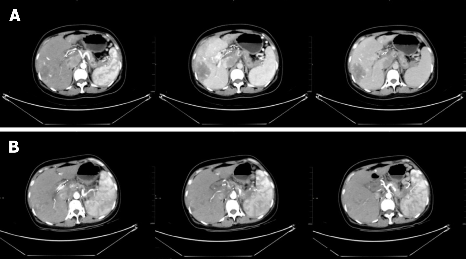
Figure 4 Imaging findings.
A: Abdominal enhanced computed tomography (CT) in February 2020 demonstrated the following: (1) The presence of multiple intrahepatic tumors. Compared with the previous image (November 2019), the lesion volume of the hepatic hilar was increased, abdominal exudation and liver injury were improved, necrosis appeared in some lesions, and little change was seen in the remaining lesions; and (2) the volume of emboli in the portal vein and inferior vena cava was increased, and varicose veins were present in the lower esophagus and gastric fundus; B: Abdominal contrast-enhanced CT in June 2020 revealed multiple tumors in the liver accompanied by the accumulation of lipiodol, and increased accumulation of lipiodol in the lesion compared with the previous lesion (February 2020). The lesion scope of the pancreatic tail was reduced.
- Citation: Gao LP, Kong GX, Wang X, Ma HM, Ding FF, Li TD. Pancreatic neuroendocrine carcinoma in a pregnant woman: A case report and review of the literature . World J Clin Cases 2021; 9(17): 4327-4335
- URL: https://www.wjgnet.com/2307-8960/full/v9/i17/4327.htm
- DOI: https://dx.doi.org/10.12998/wjcc.v9.i17.4327


