Copyright
©The Author(s) 2021.
World J Clin Cases. Jun 16, 2021; 9(17): 4318-4326
Published online Jun 16, 2021. doi: 10.12998/wjcc.v9.i17.4318
Published online Jun 16, 2021. doi: 10.12998/wjcc.v9.i17.4318
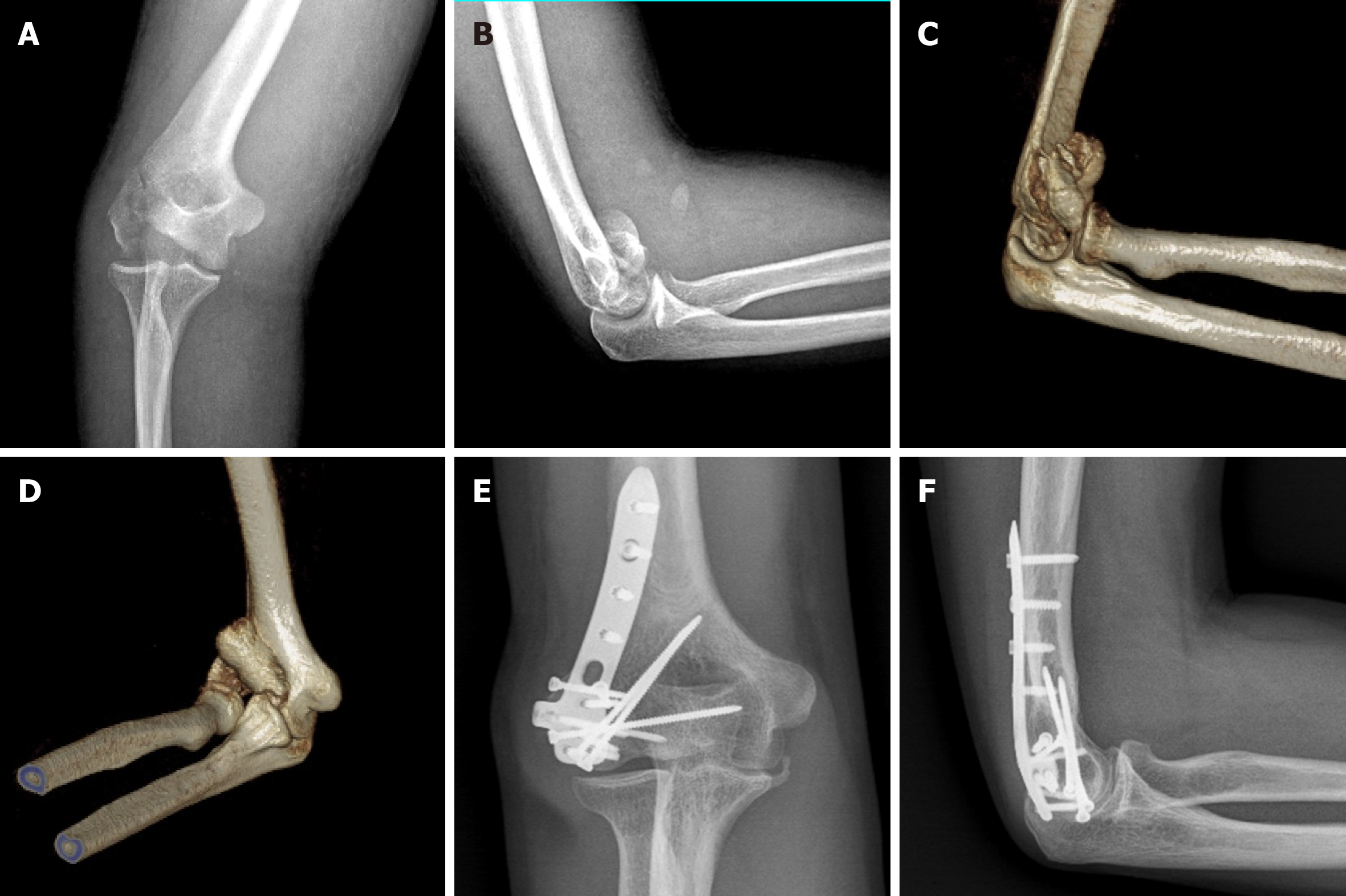
Figure 3 Imaging examinations performed before and 16 mo after surgery.
A-D: Preoperative radiographs and computed tomography scan showed a type Dubberley 2B fracture accompanied by a lateral epicondyle fracture; E and F: Radiographs taken 16 mo after surgery showed union without osteonecrosis.
- Citation: Li J, Martin VT, Su ZW, Li DT, Zhai QY, Yu B. Lateral epicondyle osteotomy approach for coronal shear fractures of the distal humerus: Report of three cases and review of the literature. World J Clin Cases 2021; 9(17): 4318-4326
- URL: https://www.wjgnet.com/2307-8960/full/v9/i17/4318.htm
- DOI: https://dx.doi.org/10.12998/wjcc.v9.i17.4318


