Copyright
©The Author(s) 2021.
World J Clin Cases. Jun 16, 2021; 9(17): 4230-4237
Published online Jun 16, 2021. doi: 10.12998/wjcc.v9.i17.4230
Published online Jun 16, 2021. doi: 10.12998/wjcc.v9.i17.4230
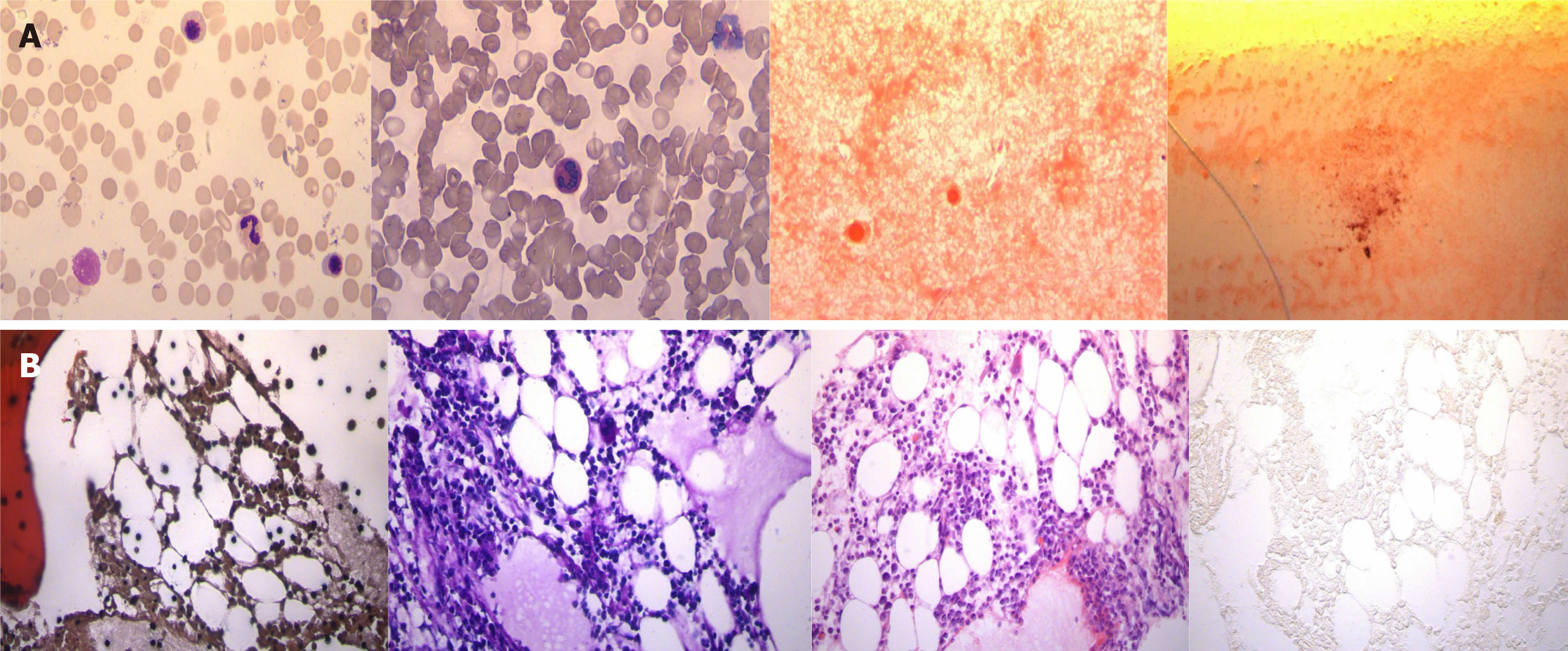
Figure 3 The results of the bone marrow biopsy.
A: Results of bone marrow aspiration-myelogram. The myelogram showed low myelodysplasia (G = 52.0%, E = 25.0%, G/E = 2.1/1); cells in the lower and middle granulocyte stages were observed, with a low proportion of mesoblastic granulocytes and a high proportion of lobulated nuclei, with obvious abnormal morphology. Cells in the lower erythroid stage could be seen with a higher proportion of late immature red cells, smaller cell bodies, different sizes of mature red cells, and some hollow enlargement. No obvious abnormality was found in lymphocytes; only one naked nucleus was found in the whole sample, with few platelets and no parasites; B: Bone marrow biopsy showed focal hyperplasia in some areas (70%) and normal hyperplasia in some areas (40%). The proportion of granulocyte red staining was generally normal. Cells at various granulocyte stages were visible, mainly in the middle and late juvenile stage; there were many megakaryocytes, mainly with lobulated nuclei, and some megakaryocytes were less abundant. Reticular fiber staining: Mf0 grade iron staining: Negative.
- Citation: Zhou XS, Lu YY, Gao YF, Shao W, Yao J. Bone marrow inhibition induced by azathioprine in a patient without mutation in the thiopurine S-methyltransferase pathogenic site: A case report. World J Clin Cases 2021; 9(17): 4230-4237
- URL: https://www.wjgnet.com/2307-8960/full/v9/i17/4230.htm
- DOI: https://dx.doi.org/10.12998/wjcc.v9.i17.4230


