
















©The Author(s) 2021.
World J Clin Cases. Jun 6, 2021; 9(16): 4052-4062
Published online Jun 6, 2021. doi: 10.12998/wjcc.v9.i16.4052
Published online Jun 6, 2021. doi: 10.12998/wjcc.v9.i16.4052
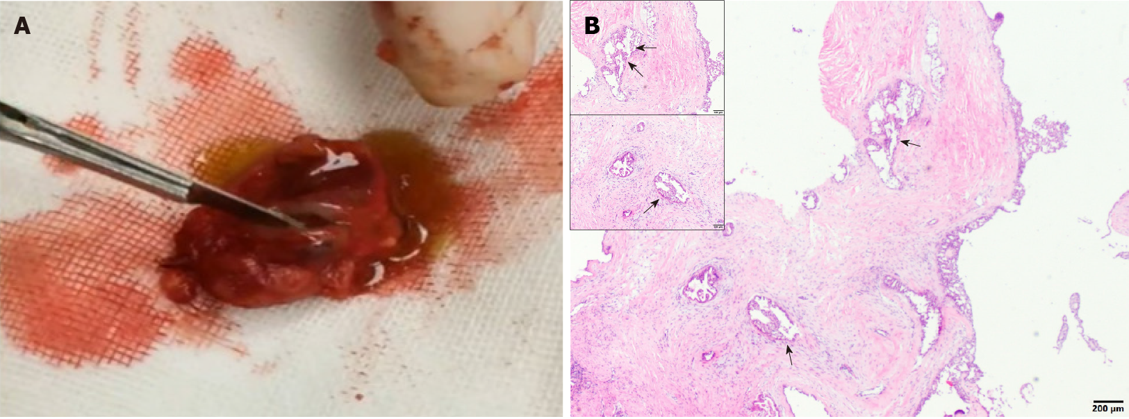
Figure 2 Clinical and histological examinations of the tumor tissues.
A: A section of the tumor, showing a solid, brownish red, cystic, mass with yellowish green liquid; B: The histological examination of the lesion showing the cystic components, areas of the cyst wall epithelium, and papillary hyperplasia to the cyst cavity (indicated using black arrows).
- Citation: Min FH, Li J, Tao BQ, Liu HM, Yang ZJ, Chang L, Li YY, Liu YK, Qin YW, Liu WW. Parotid mammary analogue secretory carcinoma: A case report and review of literature. World J Clin Cases 2021; 9(16): 4052-4062
- URL: https://www.wjgnet.com/2307-8960/full/v9/i16/4052.htm
- DOI: https://dx.doi.org/10.12998/wjcc.v9.i16.4052