Copyright
©The Author(s) 2021.
World J Clin Cases. Jun 6, 2021; 9(16): 3943-3950
Published online Jun 6, 2021. doi: 10.12998/wjcc.v9.i16.3943
Published online Jun 6, 2021. doi: 10.12998/wjcc.v9.i16.3943
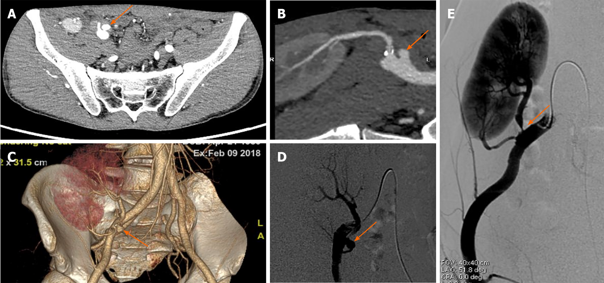
Figure 3 Computed tomography angiography and digital subtraction angiography manifestations of the extra-renal pseudo-aneurysm.
A-C: Extra-renal pseudo-aneurysm (EPSA at the anastomotic site was seen by computed tomography angiography examination (orange arrows); D and E: Severe stenosis at the proximal site of the main renal artery trunk (E) with EPSA at the anastomosis with the external iliac artery (D) was confirmed by digital subtraction angiography examination (orange arrows).
- Citation: Xu RF, He EH, Yi ZX, Li L, Lin J, Qian LX. Diagnosis and spontaneous healing of asymptomatic renal allograft extra-renal pseudo-aneurysm: A case report. World J Clin Cases 2021; 9(16): 3943-3950
- URL: https://www.wjgnet.com/2307-8960/full/v9/i16/3943.htm
- DOI: https://dx.doi.org/10.12998/wjcc.v9.i16.3943


