Copyright
©The Author(s) 2021.
World J Clin Cases. Jun 6, 2021; 9(16): 3943-3950
Published online Jun 6, 2021. doi: 10.12998/wjcc.v9.i16.3943
Published online Jun 6, 2021. doi: 10.12998/wjcc.v9.i16.3943
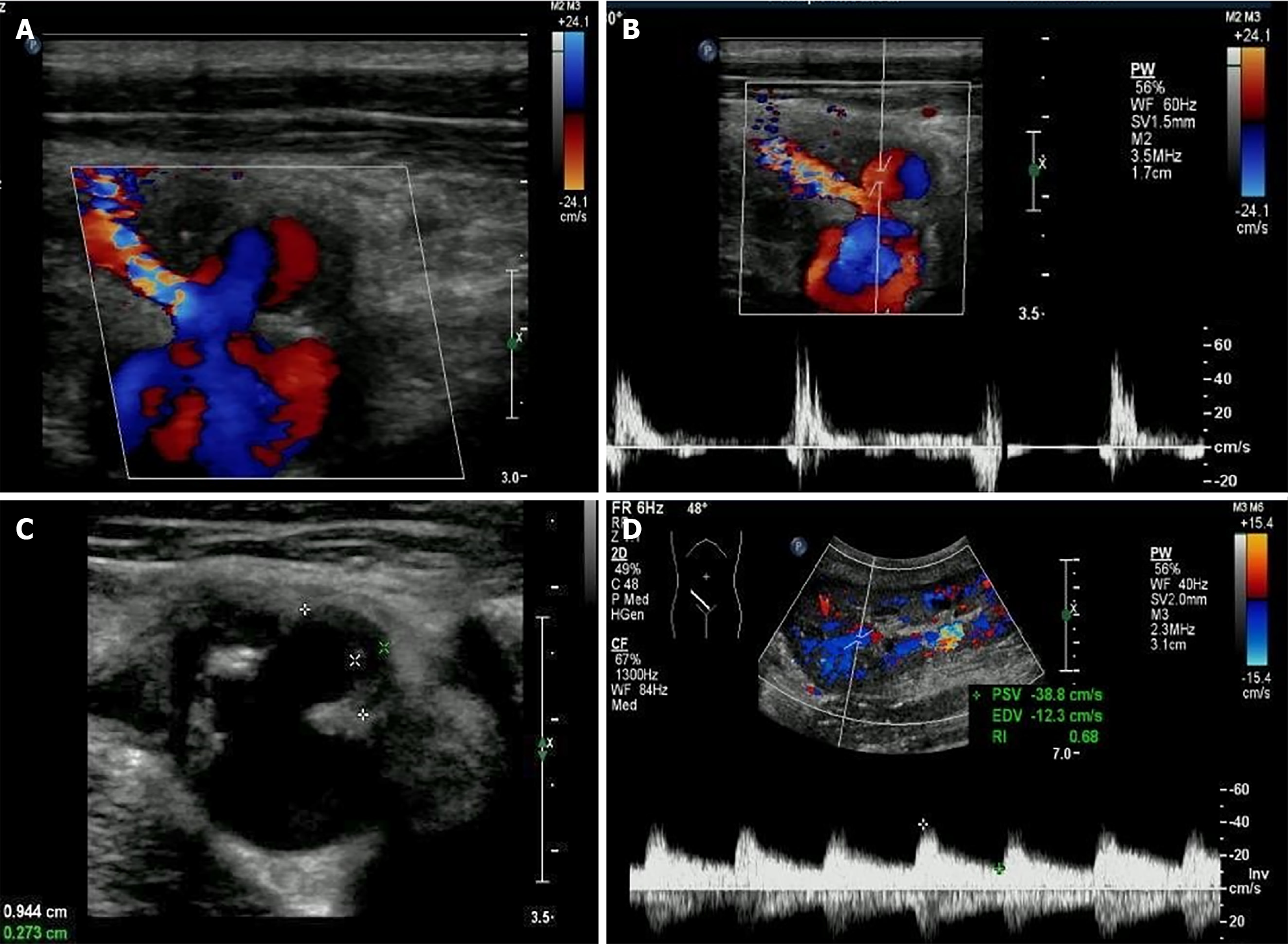
Figure 2 Extra-renal pseudo-aneurysm under ultrasonography.
A: Blood flow signal showing as swirling pattern was visible; B: To-and-fro disorganized arterial spectrum was detected with pulsed-wave Doppler; C: Hypo-echoic thrombus with thickness of 0.3 cm was observed inside the cystic structure; D: Prolonged acceleration time was longer than 70 ms of the related area inside the graft.
- Citation: Xu RF, He EH, Yi ZX, Li L, Lin J, Qian LX. Diagnosis and spontaneous healing of asymptomatic renal allograft extra-renal pseudo-aneurysm: A case report. World J Clin Cases 2021; 9(16): 3943-3950
- URL: https://www.wjgnet.com/2307-8960/full/v9/i16/3943.htm
- DOI: https://dx.doi.org/10.12998/wjcc.v9.i16.3943


