Copyright
©The Author(s) 2021.
World J Clin Cases. Jun 6, 2021; 9(16): 3943-3950
Published online Jun 6, 2021. doi: 10.12998/wjcc.v9.i16.3943
Published online Jun 6, 2021. doi: 10.12998/wjcc.v9.i16.3943
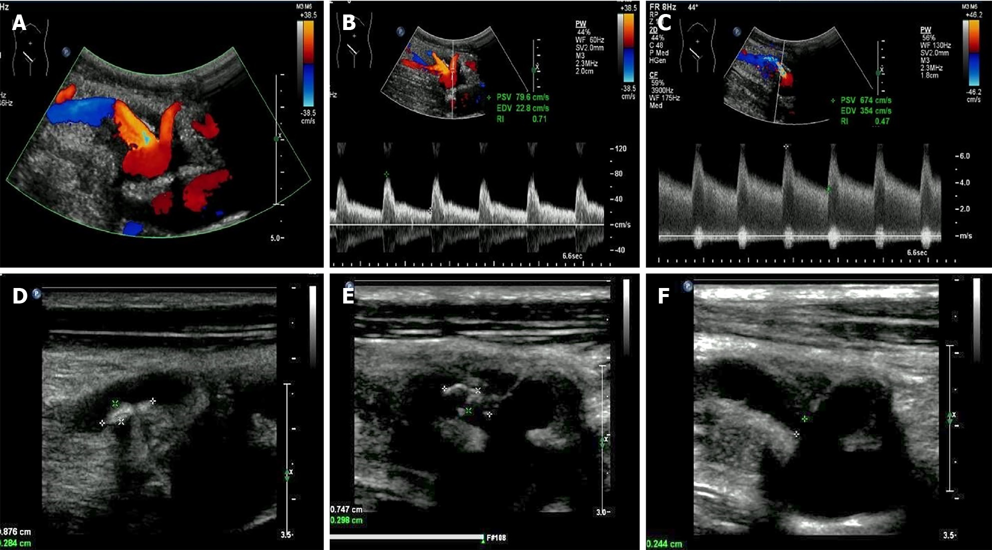
Figure 1 Ultrasonographic examination.
A: Two artery trunks anastomosed with the side of the external iliac artery were detected; B: Peak systolic velocity (PSV) of the low pole artery was 79.6 cm/s; C: PSV of 674 cm/s with color aliasing at the proximal site of the main renal trunk was detected; D and E: Multiple hypo-echoic plaques attached to the wall of the main renal artery trunk was detected; F: The residual lumen at the proximal site of the main renal artery trunk was 0.2 cm in diameter.
- Citation: Xu RF, He EH, Yi ZX, Li L, Lin J, Qian LX. Diagnosis and spontaneous healing of asymptomatic renal allograft extra-renal pseudo-aneurysm: A case report. World J Clin Cases 2021; 9(16): 3943-3950
- URL: https://www.wjgnet.com/2307-8960/full/v9/i16/3943.htm
- DOI: https://dx.doi.org/10.12998/wjcc.v9.i16.3943


