Copyright
©The Author(s) 2021.
World J Clin Cases. Jun 6, 2021; 9(16): 3927-3935
Published online Jun 6, 2021. doi: 10.12998/wjcc.v9.i16.3927
Published online Jun 6, 2021. doi: 10.12998/wjcc.v9.i16.3927
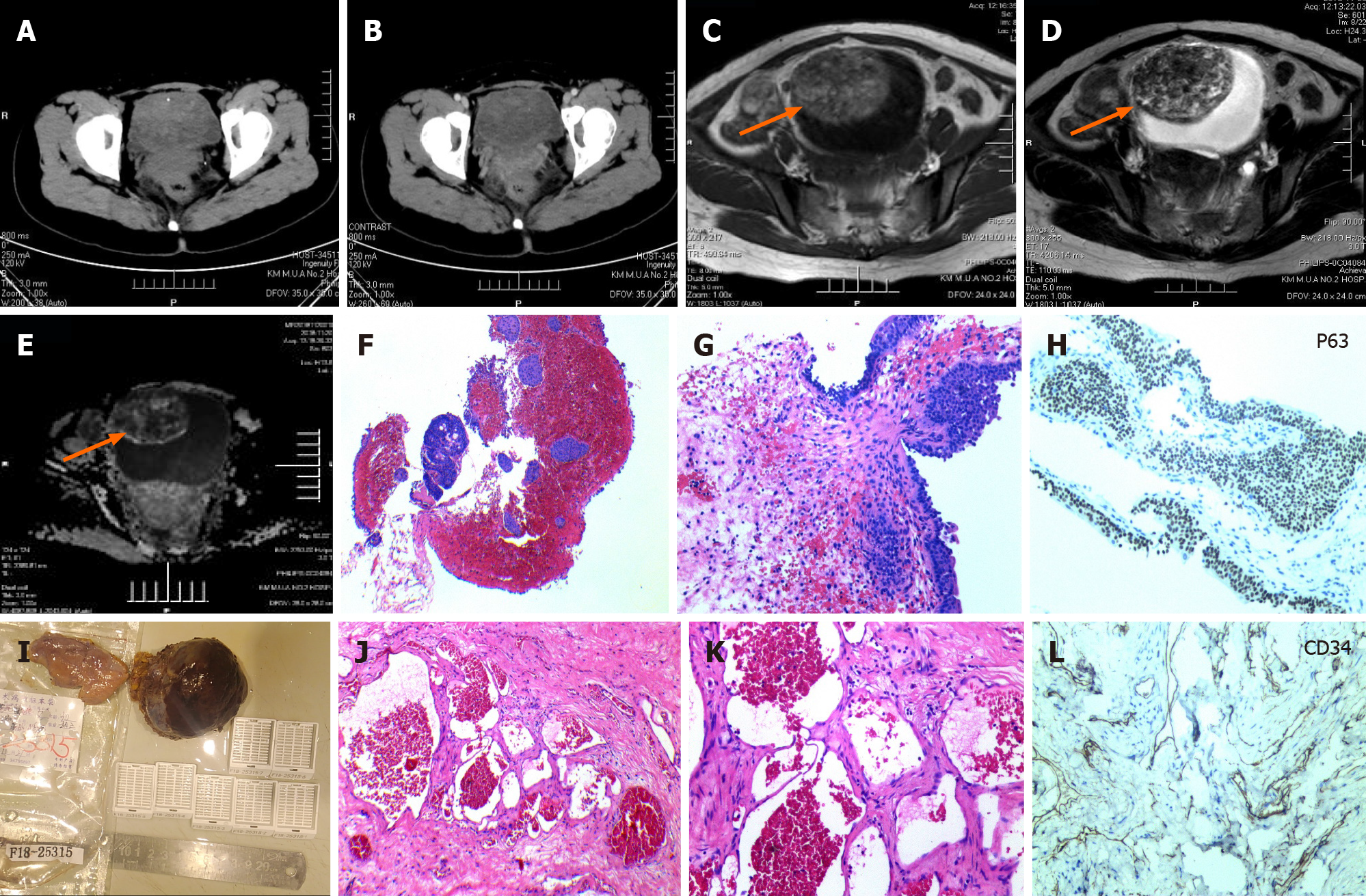
Figure 2 Computed tomography scanning, magnetic resonance imaging scanning, cystoscopy and biopsy, and pathological analysis of case 2.
A and B: Preoperative computed tomography scan image showed that the bladder wall was thickened, and patchy soft tissue density shadows and punctate calcifications could be seen in the cavity (A) along with uneven enhancement (B); C-E: Preoperative pelvic magnetic resonance imaging. The tumour was a large 6.2 cm × 6.9 cm × 5.2 cm sharply defined lesion on the right anterior and upper bladder wall, which showed intermediate signal intensity on T1-weighted images (C), heterogeneous signal intensity with a predominance of hyperintensity on T2-weighted images, and marked enhancement of the lesion (D). Diffusion weighted imaging showed a slightly higher signal, Apparent Diffusion Coefficient (ADC) showed a slightly lower signal, and the enhancement was slight (E). F and H: Cystoscopy and biopsy. The initial pathological report of haematoxylin and eosin showed gland cystitis and a single-layer flat endothelium with no nuclear atypia (F: 40 ×, G: 100 ×); endothelial cells were P63-positive (H: 100 ×); I: A specimen of the resected tumour: The gross histopathology revealed a well-circumscribed tumour bearing a vesicle-like shape with a size of 7.0 cm × 5.0 cm × 4.0 cm and a grey-brown cut surface; and J-L: Histological and immunohistochemical characteristics after partial cystectomy. Haematoxylin and eosin staining showed that the tissue structure was predominantly formed by large and dilated vessels that were engorged with blood and covered with a thin wall (J: 40 ×) but with no clear atypia of endothelial cells (K: 100 ×). Endothelial cells were CD34-positive (L: 100 ×).
- Citation: Zhao GC, Ke CX. Haemangiomas in the urinary bladder: Two case reports. World J Clin Cases 2021; 9(16): 3927-3935
- URL: https://www.wjgnet.com/2307-8960/full/v9/i16/3927.htm
- DOI: https://dx.doi.org/10.12998/wjcc.v9.i16.3927


