Copyright
©The Author(s) 2021.
World J Clin Cases. Jun 6, 2021; 9(16): 3869-3879
Published online Jun 6, 2021. doi: 10.12998/wjcc.v9.i16.3869
Published online Jun 6, 2021. doi: 10.12998/wjcc.v9.i16.3869
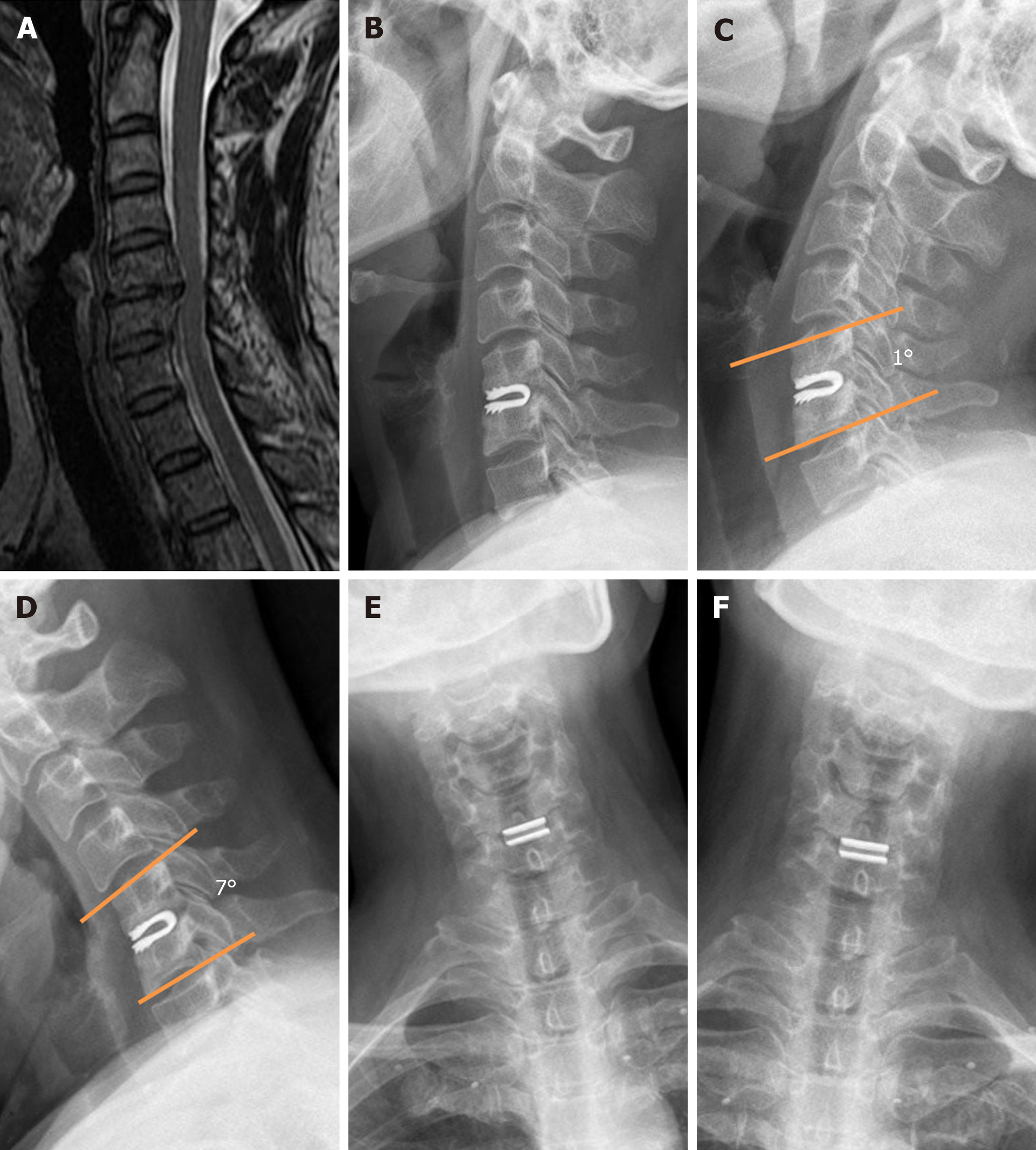
Figure 2 A 50-year-old woman suffering from C5-6 degenerative disc disease.
A: Pre-operative magnetic resonance imaging demonstrated C5-6 disc herniation; B: At the 5-year follow-up, the neutral lateral radiograph showed a normal position of dynamic cervical implant; C and D: Dynamic radiographs showing that the functional spinal unit range of motion maintained; E and F: The lateral bending at the operated level was limited.
- Citation: Zou L, Rong X, Liu XJ, Liu H. Clinical and radiological outcomes of dynamic cervical implant arthroplasty: A 5-year follow-up. World J Clin Cases 2021; 9(16): 3869-3879
- URL: https://www.wjgnet.com/2307-8960/full/v9/i16/3869.htm
- DOI: https://dx.doi.org/10.12998/wjcc.v9.i16.3869


