Copyright
©The Author(s) 2021.
World J Clin Cases. May 26, 2021; 9(15): 3637-3643
Published online May 26, 2021. doi: 10.12998/wjcc.v9.i15.3637
Published online May 26, 2021. doi: 10.12998/wjcc.v9.i15.3637
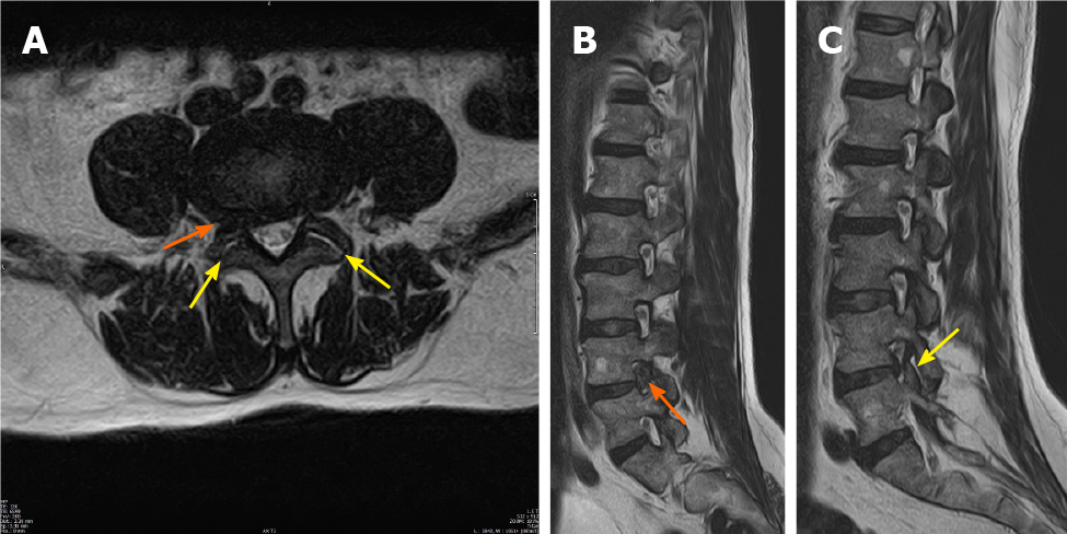
Figure 1 Magnetic resonance imaging scan done 2 d before the last infiltration, 1.
5 mo preoperatively. A: T2-weighted axial magnetic resonance imaging (MRI) image; B: T2-weighted right-sided sagittal MRI image showing right-sided foraminal lumbar disc herniation at L4-L5 (orange arrows), with compression of the right-sided exiting L4-nerve root; C: T2-weighted left-sided sagittal MRI image showing a slight bulging of the L4-L5 disc, without compression of the exiting nerve root. In (A) and (C) fluid collections can be seen in both facet joints (yellow arrows), without visible signs of edema.
- Citation: Kerckhove MFV, Fiere V, Vieira TD, Bahroun S, Szadkowski M, d'Astorg H. Postoperative pain due to an occult spinal infection: A case report. World J Clin Cases 2021; 9(15): 3637-3643
- URL: https://www.wjgnet.com/2307-8960/full/v9/i15/3637.htm
- DOI: https://dx.doi.org/10.12998/wjcc.v9.i15.3637


