Copyright
©The Author(s) 2021.
World J Clin Cases. May 26, 2021; 9(15): 3623-3630
Published online May 26, 2021. doi: 10.12998/wjcc.v9.i15.3623
Published online May 26, 2021. doi: 10.12998/wjcc.v9.i15.3623
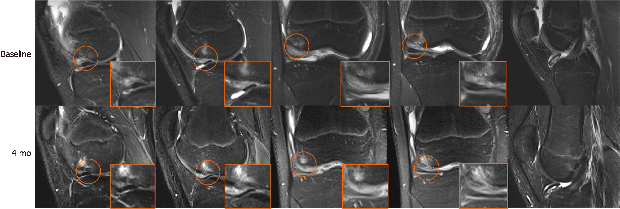
Figure 1 Sequential coronal and sagittal T2-weighted magnetic resonance imaging images at baseline and 4 mo.
The focal subchondral bone and cartilage defect (orange circle) improved progressively over time, and the whole-organ magnetic resonance imaging score improved from 25 at baseline to 14 at 4 mo. Initial integration was observed between regenerative and native subchondral bone and cartilage. The loose body still existed.
- Citation: Zhang SY, Xu HH, Xiao MM, Zhang JJ, Mao Q, He BJ, Tong PJ. Subchondral bone as a novel target for regenerative therapy of osteochondritis dissecans: A case report. World J Clin Cases 2021; 9(15): 3623-3630
- URL: https://www.wjgnet.com/2307-8960/full/v9/i15/3623.htm
- DOI: https://dx.doi.org/10.12998/wjcc.v9.i15.3623


